. 2017 Aug 1;7(1):6979.
doi: 10.1038/s41598-017-07535-8.

PRR2, a pseudo-response regulator, promotes salicylic acid and camalexin accumulation during plant immunity

Affiliations

PRR2, a pseudo-response regulator, promotes salicylic acid and camalexin accumulation during plant immunity

C Cheval et al. Sci Rep. .

Abstract

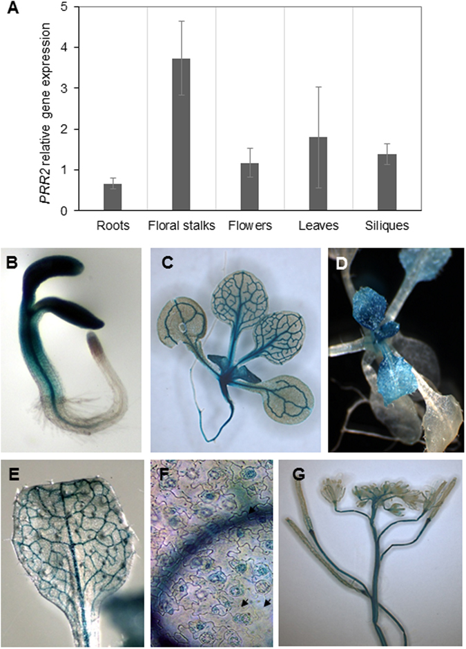
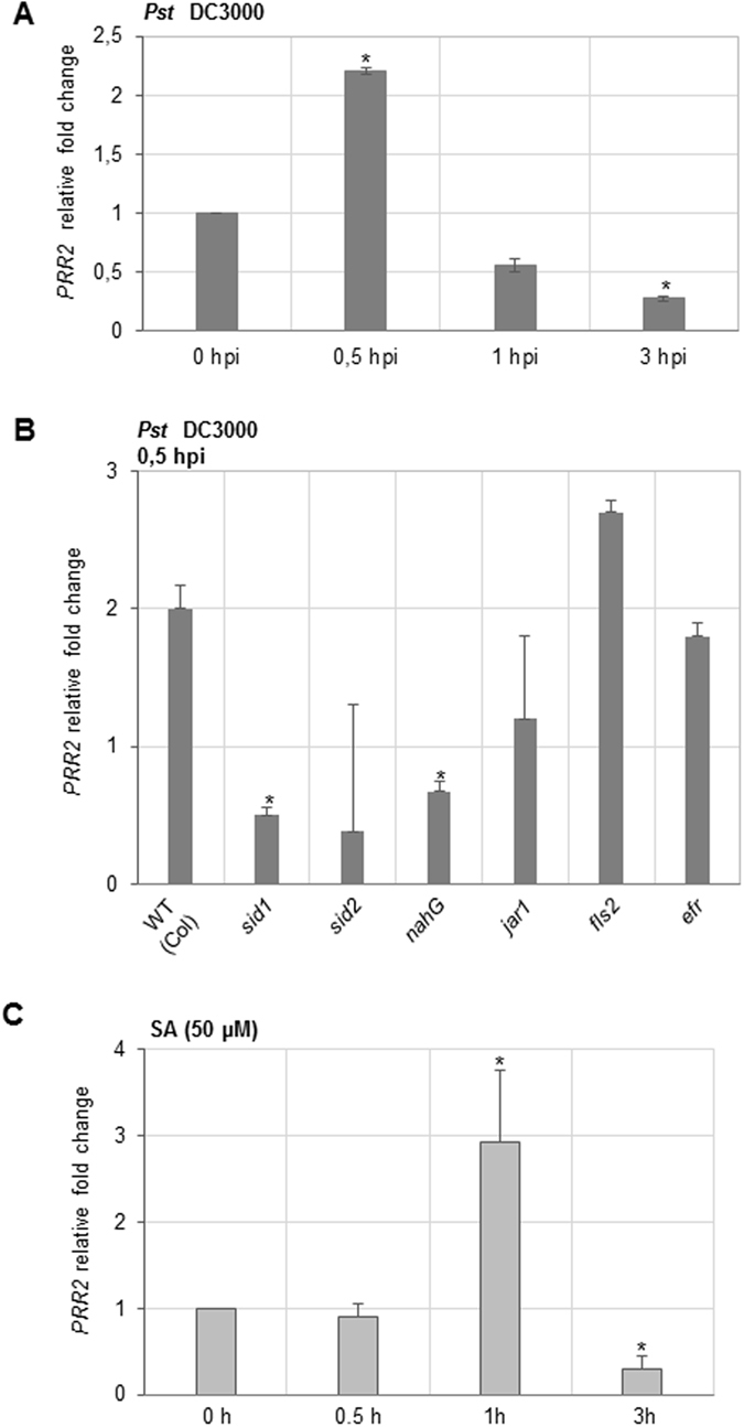
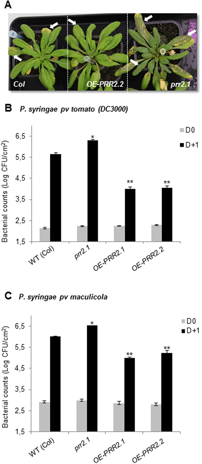
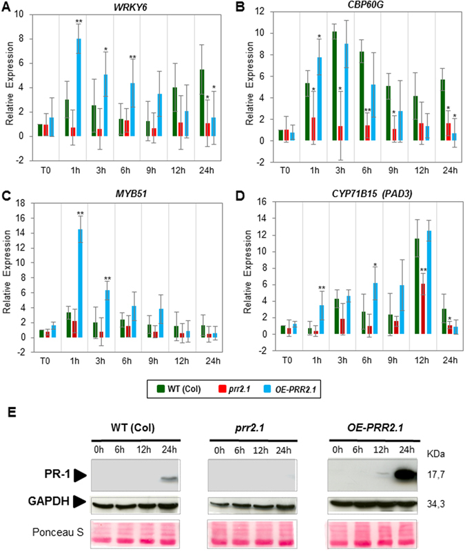
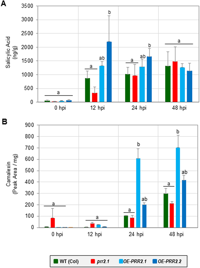
Calcium signalling mediated by Calmodulin (CaM) and calmodulin-like (CML) proteins is critical to plant immunity. CaM and CML regulate a wide range of target proteins and cellular responses. While many CaM-binding proteins have been identified, few have been characterized for their specific role in plant immunity. Here, we report new data on the biological function of a CML-interacting partner, PRR2 (PSEUDO-RESPONSE REGULATOR 2), a plant specific transcription factor. Until now, the physiological relevance of PRR2 remained largely unknown. Using a reverse genetic strategy in A. thaliana, we identified PRR2 as a positive regulator of plant immunity. We propose that PRR2 contributes to salicylic acid (SA)-dependent responses when challenged with the phytopathogenic bacterium Pseudomonas syringae. PRR2 is transcriptionally upregulated by SA and P. syringae, enhances SA biosynthesis and SA signalling responses; e.g. in response to P. syringae, PRR2 induces the production of SA and the accumulation of the defence-related protein PR1. Moreover, PRR2 overexpressing lines exhibit an enhanced production of camalexin, a phytoalexin that confers enhanced resistance against pathogens. Together, these data reveal the importance of PRR2 in plant immune responses against P. syringae and suggest a novel function for this particular plant specific transcription factor in plant physiology.


Conflict of interest statement

The authors declare that they have no competing interests.

Figures

Figure 1
Figure 1
PRR2 gene expression patterns analyses. (A) RT-QPCR analysis of PRR2 transcript levels in different organs of Arabidopsis thaliana (Col) plants. Total RNAs purified from different tissues of plants grown under long-day conditions were subjected to RT-QPCR with specific primers for PRR2. The data were obtained by the comparative 2−ΔΔCT method using actin8 as a reference gene. The illustrated values are means ± SD of four independent experiments. (B to G) Histochemical GUS staining in tissues of transgenic plants transformed with PRR2 promoter::uidA reporter construct. (B) 4-day-old seedling; (C) 16-day-old seedling; (D) 3-week-old plant; (E) adult leaf; (F) leaf epidermal tissue and guard cells (arrows); (G) floral stalk and developing siliques. T3 generation plants were grown under long-day conditions and subjected to GUS staining overnight. The observations illustrated are representative of patterns obtained with five independent transgenic lines.
Figure 2
Figure 2
PRR2 gene expression analyses in response to Pseudomonas syringae inoculation and to salicylic acid treatment. (A) PRR2 gene expression in response to P. syringae infection. Leaves of 4-week-old Col plants were inoculated with Pst DC3000 at 5.107 cfu.mL−1. Samples were collected at 0, 0.5, 1 and 3 h post-inoculation (hpi). (B) PRR2 gene expression in mutants defective for hormonal production and MAMP perception. PRR2 expression was monitored 0.5 h after Pst DC3000 inoculation in 4-week-old Arabidopsis mutants defective for SA production (sid1, sid2 and a line carrying the nahG transgene), JA (jar1) signalling pathways but also in mutants altered in flagellin (fls2) or EF-Tu (efr) perception. (C) Time-course of PRR2 gene expression in response to exogenous application of salicylic acid (SA 50 µM). Seedlings were collected at 0, 0.5, 1 and 3 h after SA treatment. All the expression analyses are presented as a fold change relative to mock treatment. Relative transcript quantification was assayed by quantitative real-time PCR and calculated by the comparative 2−ΔΔCT method using actin8 as a reference gene. Data illustrated represent the mean ± SE of three biological replicates from two independent experiments. Asterisks (*) above histograms (ANOVA, p-value 0.05) indicate significant changes of PRR2 gene expression in these different genetic backgrounds compared to WT (Col).
Figure 3
Figure 3
Altered susceptibility to Pseudomonas syringae in prr2 knock-down mutants and in transgenic lines overexpressing PRR2. (A) Disease symptoms observed in 4-week-old Arabidopsis leaves (WT (Col), OE-PRR2.2 and prr2.1) caused by Pst DC3000 infection. Leaves were sprayed with 5.107 cfu.mL−1 of Pst DC3000 and pictures were taken 7 days post-infection. Arrows indicate leaves exhibiting pronounced chlorosis symptoms. (B and C) Quantification of in planta bacterial growth were performed at 0 and 1 dpi with Pst DC3000 (B) or Pseudomonas syringae pv maculicola (C) in WT (Col), prr2.1 mutant and over-expressing transgenic lines OE-PRR2.1 and OE-PRR2.2. Data are representatives of 8 replicates from three independent experiments (n = 24). Error bars indicate SE. P values were calculated using the two-tailed Mann-Whitney U-test to indicate significant differences in bacterial growth in these different genetic backgrounds compared to WT (*p < 0.05, **p < 0.01).
Figure 4
Figure 4
Expression analysis of defence marker genes and detection of PR1 protein in prr2 knock-down mutants and in transgenic line overexpressing PRR2. (A,B,C and D) Analyses of WRKY6 (A), CBP60G (B), MYB51 (C), CYP71B15/PAD3 (D) marker genes in different prr2 genetic backgrounds. Leaves of 4-week-old Arabidopsis WT (Col), mutant prr2.1, and OE-PRR2.1 were inoculated with 5 × 107 cfu.mL−1 of Pst DC3000 and harvested at 0, 1, 3, 6, 9, 12 and 24 hours post-inoculation (hpi). The fold changes relative to the mock treatment were determined by RT-Q PCR. The values are means ± standard deviation of three independent experiments. Asterisks (*) above histograms indicate significant changes of PRR2 gene expression in these different genetic backgrounds compared to WT (Col) (student t-test with p-value < 0.05 (*) or p-value 0.01 (**)). (E) Detection of PR1 by immunoblot experiment in WT plants and prr2 genotypes. PR1 accumulation was detected in leaves of 3-week-old plants at 0, 6, 12, 24 hpi after spraying with Pst DC3000 (2 × 108 cfu.mL−1). Equal loading was confirmed by immunoblot detection of GAPDH and Ponceau S staining of the membrane (middle and lower panels). The illustrated blot is representative of three biological replicates.
Figure 5
Figure 5
Quantification of total SA and camalexin content in prr2 genotypes in response to Pseudomonas syringae inoculation. (A) Quantification of SA levels in leaves of 4-week-old plants of Arabidopsis thaliana in control conditions or after inoculation with Pst DC3000. SA quantifications were performed at 0 h, 12 h, 24 h and 48 h post-inoculation in WT (Col) and prr2 plants (prr2.1, OE-PRR2.1 and OE-PRR2.2) with Pst DC3000 at 107 cfu.mL−1. (B) Quantification of camalexin levels in leaves of 4-week-old plants of Arabidopsis thaliana in control conditions or after inoculation with Pst DC3000. Camalexin content was measured in WT, prr2 and OE-PRR2 lines after inoculation with Pst DC3000. For A and B, each bar represents the mean and standard error of three biological replicates. Bars sharing the same letter are not significantly different according to Dunnett’s test (p-value < 0.05).

References

    1. Tsuda K, Somssich IE. Transcriptional networks in plant immunity. New Phytol. 2015;206:932–947. doi: 10.1111/nph.13286. - DOI - PubMed
    1. Nakashima K, Ito Y, Yamaguchi-Shinozaki K. Transcriptional regulatory networks in response to abiotic stresses in Arabidopsis and grasses. Plant Physiol. 2009;149:88–95. doi: 10.1104/pp.108.129791. - DOI - PMC - PubMed
    1. Buscaill P, Rivas S. Transcriptional control of plant defence responses. Curr Opin Plant Biol. 2014;20C:35–46. doi: 10.1016/j.pbi.2014.04.004. - DOI - PubMed
    1. Schutze K, Harter K, Chaban C. Post-translational regulation of plant bZIP factors. Trends Plant Sci. 2008;13:247–255. doi: 10.1016/j.tplants.2008.03.002. - DOI - PubMed
    1. Bemer M, van Dijk AD, Immink RG, Angenent GC. Cross-Family Transcription Factor Interactions: An Additional Layer of Gene Regulation. Trends Plant Sci. 2017;22:66–80. doi: 10.1016/j.tplants.2016.10.007. - DOI - PubMed

Publication types

MeSH terms