. 2017 Aug 1;7(1):7072.
doi: 10.1038/s41598-017-07288-4.

Bacterial membrane vesicles transport their DNA cargo into host cells

Affiliations

Bacterial membrane vesicles transport their DNA cargo into host cells

Natalie J Bitto et al. Sci Rep. .

Abstract

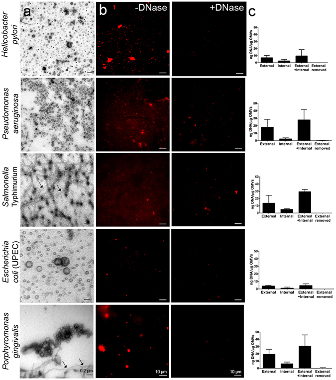
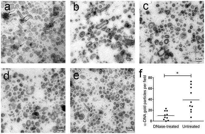
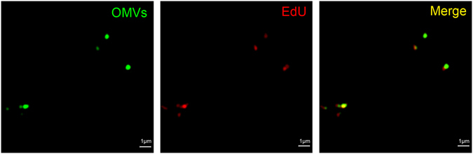
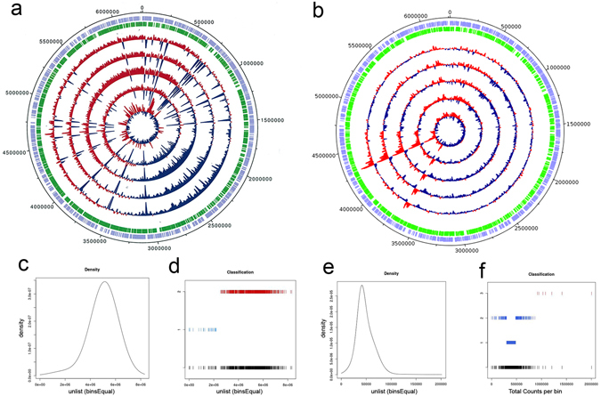
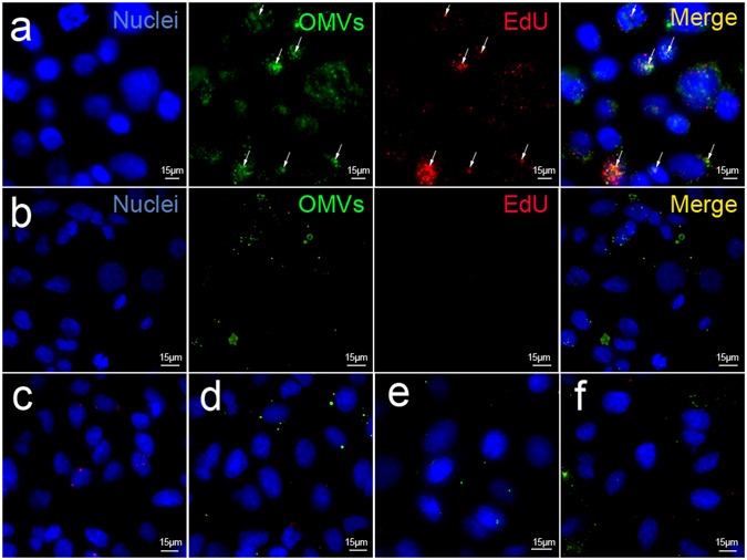
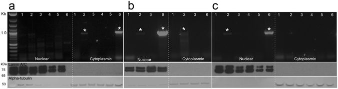
Bacterial outer membrane vesicles (OMVs) are extracellular sacs containing biologically active products, such as proteins, cell wall components and toxins. OMVs are reported to contain DNA, however, little is known about the nature of this DNA, nor whether it can be transported into host cells. Our work demonstrates that chromosomal DNA is packaged into OMVs shed by bacteria during exponential phase. Most of this DNA was present on the external surfaces of OMVs, with smaller amounts located internally. The DNA within the internal compartments of Pseudomonas aeruginosa OMVs were consistently enriched in specific regions of the bacterial chromosome, encoding proteins involved in virulence, stress response, antibiotic resistance and metabolism. Furthermore, we demonstrated that OMVs carry DNA into eukaryotic cells, and this DNA was detectable by PCR in the nuclear fraction of cells. These findings suggest a role for OMV-associated DNA in bacterial-host cell interactions and have implications for OMV-based vaccines.


Conflict of interest statement

The authors declare that they have no competing interests.

Figures

Figure 1
Figure 1
OMVs from Gram-negative bacteria carry DNA. (a) Transmission electron microscopy (TEM) of OMVs from Gram-negative bacteria (70× magnification, scale bar = 0.2 μm). Arrows indicate the presence of flagella in the S. Typhimurium and P. gingivalis OMV preparations. (b) Confocal images of OMVs treated with and without DNase and stained with the membrane permeable DNA stain SYTO-61, scale bar = 10 μm. (c) Quantification of internal and external OMV-associated DNA by Quant-iT PicoGreen dsDNA assay (n = 3). Complete removal of external DNA was verified by quantifying the DNA on intact DNase-treated OMVs (external removed).
Figure 2
Figure 2
P. aeruginosa OMVs are associated with DNA external and internal to the vesicle membrane. TEM of (a) Untreated OMVs labeled with anti-DNA antibody. Both internal and external OMV-associated DNA is observed (indicated by arrows). (b) DNase-treated OMVs labeled with anti-DNA antibody showing internal OMV-associated DNA only. (c) DNase treatment post-sectioning to remove exposed internal OMV-associated DNA, followed by detection with anti-DNase antibody. (d) Secondary antibody and protein-A-gold (PAG) only and (e) PAG only. Scale bar = 0.2 μm; 100× magnification. (f) Quantification of α-DNA labeling of DNase-treated versus untreated OMVs (p = 0.0012, n = 10 fields of view).
Figure 3
Figure 3
Super resolution microscopy of OMVs carrying bacterial DNA. OMVs were labeled with the lipophilic stain, DiO (green), which targets the OMV membrane, while EdU-incorporated DNA (red) was labeled by a 2-step process. The first step involved incubation with 12.5 μM biotin azide, during which the azide moiety was bound specifically to the alkyne backbone of the EdU molecule in the presence of a copper catalyst. The second step involved incubation with 5 μg/ml streptavidin-conjugated 568 Alexa Fluor® to fluorescently label the biotin-azide bound to the EdU. Scale bar = 1 μm.
Figure 4
Figure 4
Analysis of total and internal OMV-derived DNA sequences from five separate biological replicates of P. aeruginosa OMVs. Circular plots for the total (a) and internal (b) DNA read count across each of the replicates show read count at each base pair position. Positions showing greater than average read density are coloured red while those with less than average density are coloured blue. A peak in read count density is visible in the internal DNA at approximately 4,290,000 bp. The outer circles show the location of the genes on the positive (purple) and negative (green) strands of the reference genome. The total read counts from 603 equally-sized and contiguous bins spanning the genome were computed and plotted as density distributions for the total (c) and internal (d) DNA. A Kolmogorov-Smirnov test revealed that the read count distribution for the total DNA did not differ from a normal distribution, whereas the internal DNA possessed a long tail at the upper end of the distribution which created a significant deviation from the normal distribution (p = 1.382 × 10−6). The Gaussian finite mixture models confirmed these observations (d,f). The counts from the majority of the total DNA bins were contained within a dominant Gaussian distribution (red), while a secondary Gaussian distribution represented a small subset of bins with relatively low read densities (blue) (d). In contrast, the counts for the internal DNA bins were represented by three Gaussian distributions: two overlapping distributions (blue) represented the majority of bins with counts ranging from 0 to approximately 800 000; and a third Gaussian distribution (red) represented bins with counts at higher values (f).
Figure 5
Figure 5
Confocal microscopy showing internalization of OMV-associated DNA in A549 lung epithelial cells. EdU-labeled OMVs, either (a) untreated or (b) DNase-treated, were labeled with the lipophilic stain DiO then added to A549 cells for 5 h, washed to remove non-internalized OMVs, fixed and permeabilized. EdU was detected in a 2-step reaction consisting of incubation with biotin azide, in which the azide binds specifically to the alkyne backbone of EdU, followed by fluorescent detection with streptavidin-conjugated Alexa-Fluor 568. Co-localization of DiO (green) and EdU (red) labels in internalized OMVs are indicated by arrows. Nuclei are stained with DAPI (blue). Merged images showing control samples in which either: (c) biotin azide and streptavidin-AlexaFluor 568 only, (d) no biotin azide or (e) DiO only were added to cells. (f) Non-permeabilized cells. Scale bar = 15 μm.
Figure 6
Figure 6
DNA derived from P. aeruginosa OMVs is detected in the nuclear fractions of epithelial cells. A549 cells were either left untreated (1), treated with intact OMVs (2), DNase-treated OMVs (3), disrupted OMVs (4), free P. aeruginosa gDNA (5), or plasmid pGL3c (6) transfected with Lipofectamine 2000 (Life Technologies). After treatment for (a) 4 h, (b) 8 h and (c) 18 h, cells were DNase-treated to remove non-internalized DNA, followed by extraction of cytoplasmic and nuclear compartments. PCR products, corresponding to OMV-derived DNA and pGL3c (859 bp and 966 bp, respectively) are indicated by asterisks. Nuclear and cytoplasmic fractions were confirmed by Western blotting using anti-lamin A/C and -alpha tubulin antibodies, respectively.

References

    1. Brown L, Wolf JM, Prados-Rosales R, Casadevall A. Through the wall: extracellular vesicles in Gram-positive bacteria, mycobacteria and fungi. Nat Rev Micro. 2015;13:620–630. doi: 10.1038/nrmicro3480. - DOI - PMC - PubMed
    1. Lee, E.-Y., Choi, D.-Y., Kim, D.-K. & Kim, J.-W. Gram-positive bacteria produce membrane vesicles: proteomics-based characterization of Staphylococcus aureus-derived membrane vesicles. Proteomics9, 5425–5436 (2009). - PubMed
    1. Deatherage, B. L. & Cookson, B. T. Membrane vesicle release in bacteria, eukaryotes, and archaea: a conserved yet underappreciated aspect of microbial life. Infect Immun80, 1948–1957 (2012). - PMC - PubMed
    1. Stoorvogel W, Kleijmeer MJ, Geuze HJ, Raposo G. The biogenesis and functions of exosomes. Traffic. 2002;3:321–330. doi: 10.1034/j.1600-0854.2002.30502.x. - DOI - PubMed
    1. Samuel M, Bleackley M, Anderson M, Mathivanan S. Extracellular vesicles including exosomes in cross kingdom regulation: a viewpoint from plant-fungal interactions. Front Plant Sci. 2015;6:766. doi: 10.3389/fpls.2015.00766. - DOI - PMC - PubMed

Publication types

LinkOut - more resources