. 2017 Aug 1;7(1):7004.
doi: 10.1038/s41598-017-06358-x.

High-Resolution Patterned Cellular Constructs by Droplet-Based 3D Printing

Affiliations

High-Resolution Patterned Cellular Constructs by Droplet-Based 3D Printing

Alexander D Graham et al. Sci Rep. .

Abstract

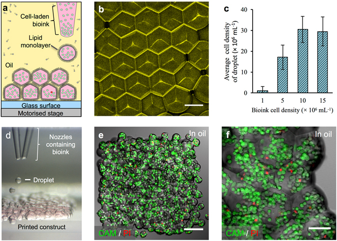
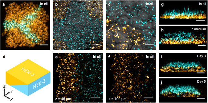
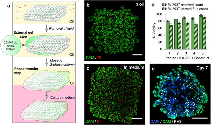
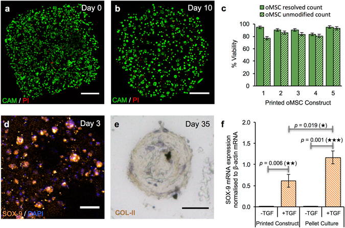
Bioprinting is an emerging technique for the fabrication of living tissues that allows cells to be arranged in predetermined three-dimensional (3D) architectures. However, to date, there are limited examples of bioprinted constructs containing multiple cell types patterned at high-resolution. Here we present a low-cost process that employs 3D printing of aqueous droplets containing mammalian cells to produce robust, patterned constructs in oil, which were reproducibly transferred to culture medium. Human embryonic kidney (HEK) cells and ovine mesenchymal stem cells (oMSCs) were printed at tissue-relevant densities (107 cells mL-1) and a high droplet resolution of 1 nL. High-resolution 3D geometries were printed with features of ≤200 μm; these included an arborised cell junction, a diagonal-plane junction and an osteochondral interface. The printed cells showed high viability (90% on average) and HEK cells within the printed structures were shown to proliferate under culture conditions. Significantly, a five-week tissue engineering study demonstrated that printed oMSCs could be differentiated down the chondrogenic lineage to generate cartilage-like structures containing type II collagen.


Conflict of interest statement

Hagan Bayley is the Founder, a Director and a shareholder of OxSyBio, a company engaged in the development of printed tissues and tissue-like materials. Work in the Bayley laboratory at the University of Oxford is supported in part by OxSyBio. During revision of the paper, Alexander Graham, Sam Olof, Madeline Burke and Stuart Box became employees of OxSyBio.

Figures

Figure 1
Figure 1
3D printing of cellular constructs. (a) Schematic of cell printing. The dispensing nozzle ejects cell-containing bioink droplets into a lipid-containing oil. The droplets are positioned by the programmed movement of the oil container. The droplets cohere through the formation of droplet interface lipid bilayers. (b) A confocal fluorescence micrograph showing droplet interface bilayers (stained yellow) within a cell-free printed construct (11 × 14 × 7 droplets). The bilayers were visualised by adding sulforhodamine-101 (~10 μM) to the print solution. (Scale bar = 100 μm). (c) Histogram showing the mean HEK-293T cell density in printed droplets under oil as a function of the cell density in the bioink. The cell density was calculated as the mean number of cells per droplet (n = 25) divided by the mean droplet volume. Error bars represent the compound error of droplet size variance and cell per droplet variance. (d) A bright-field micrograph of a patterned cell junction, containing two cell types, printed as successive layers of 1 nL droplets (d = 130 μm) ejected from two glass nozzles (d = ~150 μm). (e) A confocal fluorescence micrograph of a printed HEK-293T cellular construct (11 × 14 × 2 droplets) under oil. Live/dead cell staining was performed with calcein-AM (CAM, green) and propidium iodide (PI, red), respectively. Visible are approximately 700 cells at 4 × 107 cells mL−1 with a viability of 85% (determined by manual cell counting). (Scale bar = 150 μm). (f) A high magnification, confocal fluorescence micrograph of a live/dead assay performed on an HEK-293T cellular construct (7 × 8 × 4 droplets) printed at a starting concentration of 1.5 × 107 cells mL−1, with a mean occupancy of 38 cells per droplet equivalent to 3 × 107 cells mL−1. Visible are some of the droplet boundaries. (Scale bar = 75 μm).
Figure 2
Figure 2
High-resolution patterning of two cell types. (ac, e,f) Confocal fluorescence micrographs of printed cellular constructs in oil, immediately after printing. HEK-293 cells stained with Deep Red (DR) or Red CMPTX (RC) CellTracker™ dyes were false-coloured blue and yellow, respectively. (a) A Y-shaped structure within a square construct (8 × 9 × 4 droplets), with a mean feature width of 180 μm. (Scale bar = 200 μm). (b) A cruciform pattern of HEK-293 cells within a square construct (10 × 12 × 5 droplets). (Scale bar = 250 μm). (c) A high magnification image of the patterned HEK-293 cells in (b). (Scale bar = 100 μm). (d) A 3D model of a cuboidal cellular construct with an interface between two HEK populations (HEK 1, yellow; and HEK 2, cyan) at a diagonal in the x-z plane. (e,f) Partial cross-sections at fixed vertical positions (45 and 192 µm respectively) of a cellular construct (21 × 24 × 7 droplets) printed based according to the model in (d), showing both HEK populations. (Scale bars = 250 μm). (gj) Side-on images of lamellar constructs, comprising CellTracker™ stained HEK-293 cells before and after phase transfer. The lower, DR-stained HEK-293 cell layers (yellow) were 3 droplets thick, while, the upper, RC-stained HEK-293 cell layers (blue) were 4 droplets thick (g,h) or 3 droplets thick (i,j). Images were recorded: (g) at day 0, in oil, immediately after printing; (h) immediately after transfer to culture medium; (i) on day 3 of culture, in medium and; (j) on day 5 of culture, in medium. (Scale bar = 250 μm).
Figure 3
Figure 3
Phase transfer and culture of printed constructs containing HEK-293T cells. (a) Gel encapsulation of a printed construct and phase transfer. The printed cellular construct was gelled by standing at 4 °C for 20 to 25 min and the lipid in the oil was removed by washes with silicone oil AR20 at room temperature. The construct was then coated with a thin layer of cell-free bioink, which was gelled by standing at 4 °C for 20 to 25 min. The gelled construct was then transferred into the upper phase of an oil-culture medium two-phase system. The construct fell through the oil into the culture medium. (b) Image of a z-stack 3D reconstruction of live/dead-stained HEK-293T cells printed as a cuboid construct (7 × 8 × 4 droplets) immediately after printing under oil. The printed droplets had a mean density of 2.9 × 10⁷ cells mL−1 with a viability of 96%. (Scale bar = 200 μm). (c) Image of a z-stack 3D reconstruction of live/dead-stained printed HEK-293T cells after gel encapsulation and transfer to culture medium. (Scale bar = 200 μm). (d) Graph showing HEK-293T cell viability (including standard error of the mean) of five printed constructs at day 0 after transfer to culture medium. Viabilities were determined by using automated object counting, values of which were used either unmodified or resolved with respect to mean cell size. (e) Image of a z-stack 3D reconstruction of immunocytochemistry performed on a construct in culture medium at day 7: cell nuclei (DAPI, blue); cytoplasm of live cells (CAM, green) and; mitotic marker (phospho-histone H3 ICC, PH3, white). (Scale bar = 200 μm).
Figure 4
Figure 4
Growth and differentiation of printed oMSCs. (a,b) Image of a z-stack 3D reconstruction of live/dead-stained printed oMSCs: (a) immediately after printing and; (b) after 10 days in culture with the TGF-β3 supplement. (Scale bars = 250 μm). (c) Graph of oMSC viabilities (including standard error of the mean) for five printed constructs immediately after transfer to culture medium. Viabilities were determined by using automated object counting, values of which were used either unmodified or resolved with respect to mean cell size. (d) Confocal fluorescence micrograph of immunocytochemistry performed on a printed oMSC construct after 3 days of culture with a TGF-β3 supplement: SOX-9 (orange); nuclei (DAPI, blue); cytoplasm of live cells (CAM, calcein-AM, green). (Scale bar = 50 μm). (e) High-magnification micrograph of immunohistochemistry performed on a printed oMSC construct after 35 days of culture with TGF-β3 supplement; type II collagen (diaminobenzidine tetrahydrochloride (DAB), brown); nuclei (hematoxylin QS, blue). (Scale bar = 25 μm). (f) Digital PCR measurements of SOX-9 mRNA expression in printed oMSC constructs (n = 22) and oMSC pellet cultures (n = 24) after 7 days in chondrogenic medium with or without supplementation of TGF-β3. Each printed and pellet sample was replicated 4 to 6 times from four oMSCs sources, each extracted from a different sheep. SOX-9 expression was normalised to an endogenous β-actin control. Error bars represent standard deviations. Differences were tested by using a paired t-test, with two-tailed p values < 0.05 considered significant.

References

    1. Derby B. Printing and prototyping of tissues and scaffolds. Science. 2012;338:921–926. doi: 10.1126/science.1226340. - DOI - PubMed
    1. Murphy SV, Atala A. 3D bioprinting of tissues and organs. Nat. Biotechnol. 2014;32:773–785. doi: 10.1038/nbt.2958. - DOI - PubMed
    1. Mironov V, Kasyanov V, Markwald RR. Organ printing: from bioprinter to organ biofabrication line. Curr. Opin. Biotechnol. 2011;22:667–673. doi: 10.1016/j.copbio.2011.02.006. - DOI - PubMed
    1. Wu W, DeConinck A, Lewis JA. Omnidirectional printing of 3D microvascular networks. Adv. Mater. 2011;23:178–183. doi: 10.1002/adma.201004625. - DOI - PubMed
    1. De Coppi P, et al. Isolation of amniotic stem cell lines with potential for therapy. Nat. Biotechnol. 2007;25:100–106. doi: 10.1038/nbt1274. - DOI - PubMed

Publication types