Influence of the Respiratory Cycle on Caudal Vena Cava Diameter Measured by Sonography in Healthy Foals: A Pilot Study
- PMID: 28766820
- PMCID: PMC5598903
- DOI: 10.1111/jvim.14793
Influence of the Respiratory Cycle on Caudal Vena Cava Diameter Measured by Sonography in Healthy Foals: A Pilot Study
Abstract
Background: Intravascular volume assessment in foals is challenging. In humans, intravascular volume status is estimated by the caudal vena cava (CVC) collapsibility index (CVC-CI) defined as (CVC diameter at maximum expiration [CVCmax ] - CVC diameter at minimal inspiration [CVCmin ])/CVCmax × 100%.
Hypothesis/objectives: To determine whether the CVC could be sonographically measured in healthy foals, determine differences in CVCmax and CVCmin , and calculate inter- and intrarater variability between 2 examiners. We hypothesized that the CVC could be measured sonographically at the subxiphoid view and that there would be a difference between CVCmax and CVCmin values.
Animals: Sixty privately owned foals <1-month-old.
Methods: Prospective study. A longitudinal subxiphoid sonographic window in standing foals was used. The CVCmax and CVCmin were analyzed by a linear mixed effect model. Inter-rater agreement and intrarater variability were expressed by Bland-Altman and intraclass correlation coefficients, respectively.
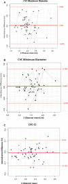
Results: Measurements were attained from 58 of 60 foals with mean age of 15 ± 7.9 days and mean weight of 75.7 ± 17.7 kg. The CVCmax was significantly different from CVCmin (D = 0.515, SE = 0.031, P < 0.001). Inter-rater agreement of the CVC-CI differed by an average of -0.9% (95% limits of agreement, -12.5 to +10.7%). Intrarater variability of CVCmax was 0.540 and 0.545, of CVCmin was 0.550 and 0.594, and of CVC-CI was 0.894 and 0.853 for observers 1 and 2, respectively.
Conclusions and clinical importance: These results indicate it is possible to reliably measure the CVC sonographically in healthy foals, and the CVC-CI may prove useful in assessing the intravascular volume status in hypovolemic foals.
Keywords: Collapsibility index; Fluid estimation; Intravascular volume status; Ultrasound.
Copyright © 2017 The Authors. Journal of Veterinary Internal Medicine published by Wiley Periodicals, Inc. on behalf of the American College of Veterinary Internal Medicine.
Figures


References
-
- Hollis AR, Boston RC, Corley KT. Plasma aldosterone, vasopressin and atrial natriuretic peptide in hypovolaemia: A preliminary comparative study of neonatal and mature horses. Equine Vet J 2008;40:64–69. - PubMed
-
- Palmer JE. Fluid therapy in the neonate: Not your mother's fluid space. Vet Clin North Am Equine Pract 2004;20:63–75. - PubMed
-
- Palmer J. Update on the management of neonatal sepsis in horses. Vet Clin North Am Equine Pract 2014;30:317–336, vii. - PubMed
-
- Stawicki SP, Braslow BM, Panebianco NL, et al. Intensivist use of hand‐carried ultrasonography to measure IVC collapsibility in estimating intravascular volume status: Correlations with CVP. J Am Coll Surg 2009;209:55–61. - PubMed
-
- Stawicki SP, Adkins EJ, Eiferman DS, et al. Prospective evaluation of intravascular volume status in critically ill patients: Does inferior vena cava collapsibility correlate with central venous pressure? J Trauma Acute Care Surg 2014;76:956–963; discussion 63‐4. - PubMed
MeSH terms
LinkOut - more resources
Full Text Sources
Other Literature Sources

