. 2017 Aug 2;18(8):1675.
doi: 10.3390/ijms18081675.

Acceleration Mechanisms of Skin Wound Healing by Autologous Micrograft in Mice

Affiliations

Acceleration Mechanisms of Skin Wound Healing by Autologous Micrograft in Mice

Shiro Jimi et al. Int J Mol Sci. .

Abstract

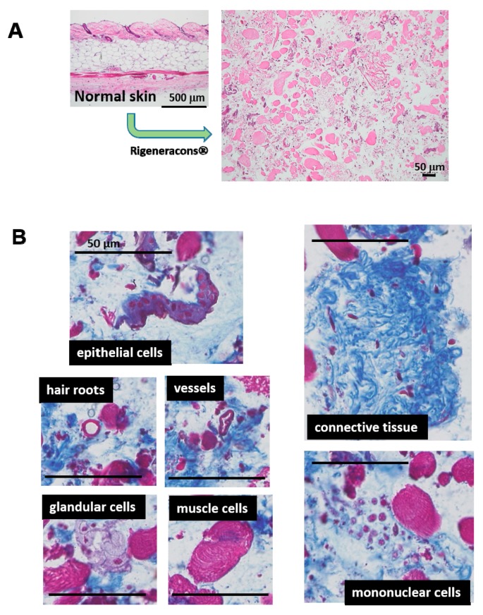
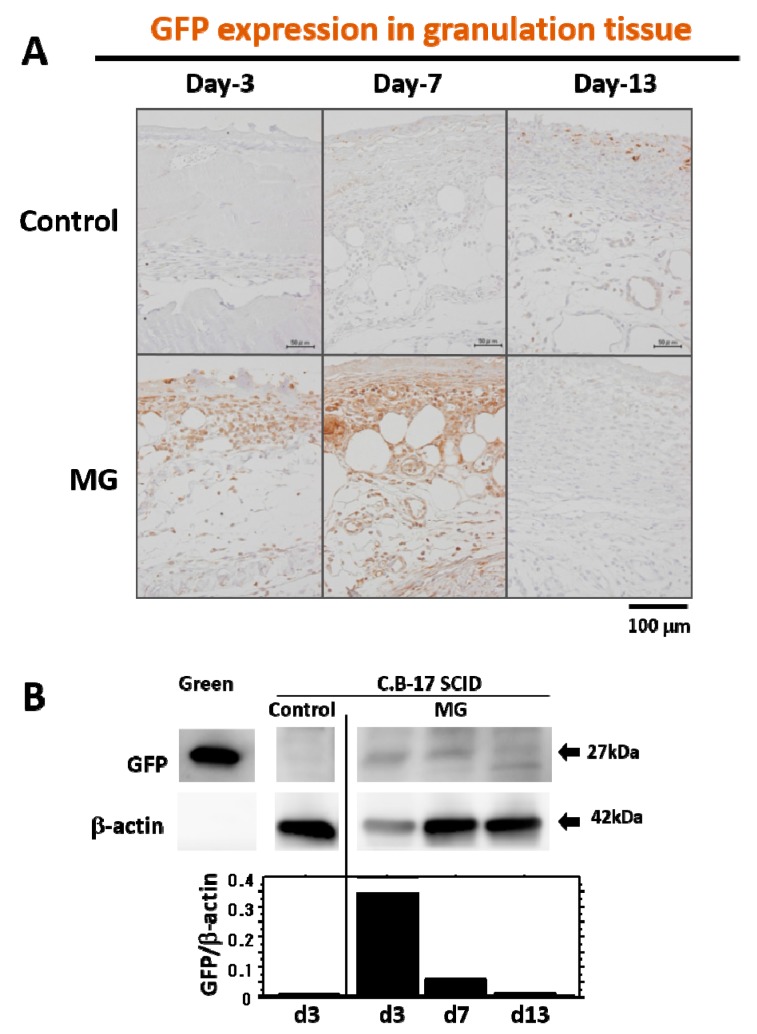
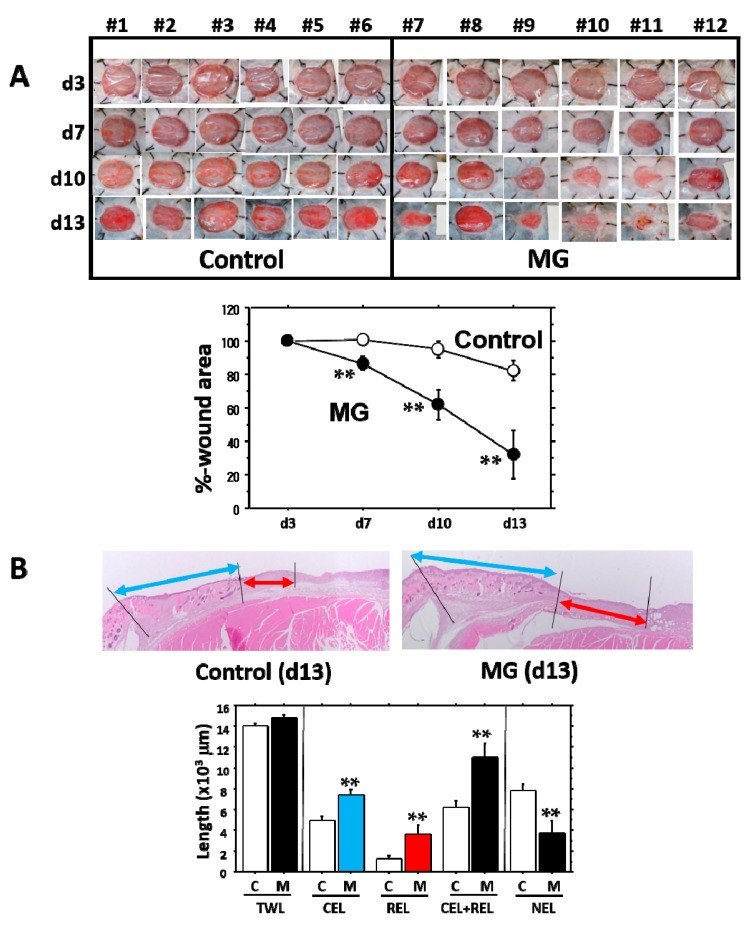
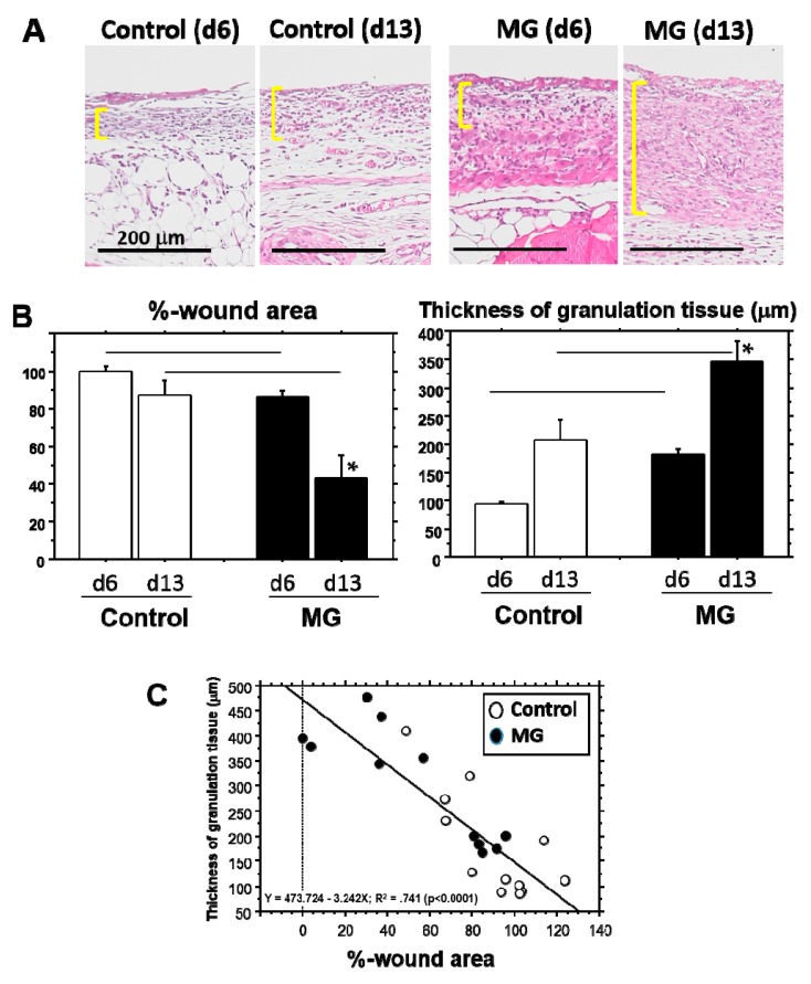
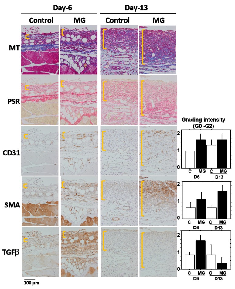
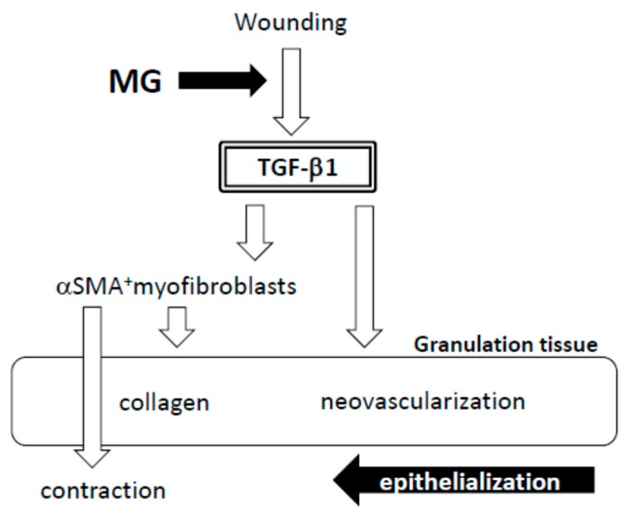
A micrograft technique, which minces tissue into micro-fragments >50 μm, has been recently developed. However, its pathophysiological mechanisms in wound healing are unclear yet. We thus performed a wound healing study using normal mice. A humanized mouse model of a skin wound with a splint was used. After total skin excision, tissue micro-fragments obtained by the Rigenera protocol were infused onto the wounds. In the cell tracing study, GFP-expressing green mice and SCID mice were used. Collagen stains including Picrosirius red (PSR) and immunohistological stains for α-smooth muscle actin (αSMA), CD31, transforming growth factor-β1 (TGF-β1) and neutrophils were evaluated for granulation tissue development. GFP-positive cells remained in granulation tissue seven days after infusion, but vanished after 13 days. Following the infusion of the tissue micrograft solution onto the wound, TGF-β1 expression was transiently upregulated in granulation tissue in the early phase. Subsequently, αSMA-expressing myofibroblasts increased in number in thickened granulation tissue with acceleration of neovascularization and collagen matrix maturation. On such granulation tissue, regenerative epithelial healing progressed, resulting in wound area reduction. Alternative alteration after the micrograft may have increased αSMA-expressing myofibroblasts in granulation tissue, which may act on collagen accumulation, neovascularization and wound contraction. All of these changes are favorable for epithelial regeneration on wound.

Keywords: TGF-β1; granulation tissue; micrograft; wound healing; αSMA.


Conflict of interest statement

The authors declare no conflict of interest.

Figures

Figure 1
Figure 1
Tissue fragments in micrografted skin solution. (A) Normal skin tissue was minced by Rigeneracons®. Obtained tissues were stained with HE and observed under a microscope; (B) Obtained tissue was also stained with Masson’s trichrome (MT), which contained all of the skin elements, including epithelial cells, hair roots, vessels, glandular cells, muscle cells and mononuclear cells. Connective tissue is also included. Scale bars = 50 μm.
Figure 2
Figure 2
GFP expression in granulation tissue after wounding. (A) Presence of GFP in granulation tissue developed after wounding was immunohistochemically detected in immune-deficient SCID mice with micrograft of skin tissue solution from GFP-positive green mouse or saline alone (control) on Days 3, 7 and 13. Photographs are representative wounds in each group. Scale bars = 100 μm; (B) Western blot analysis for GFP in granulation tissue was performed in SCID mice with micrograft of skin tissue solution from green mouse or saline alone (control) on Days 3, 7 and 13. Green mice were used as a positive control. Densitometry analysis of GFP and β-actin was also conducted; relative expression value of GFP is shown in the bar graph.
Figure 3
Figure 3
Optimal concentration of micrograft for wound healing. Optimal tissue concentration for wound healing was determined. Normal skin tissue was minced by Rigeneracons®, and its optical density at wavelengths of 450 nm/550 nm was adjusted to 1.0. The solution was serially diluted (2-, 4- and 8-times), and 200 μL of each solution were micrografted on the wound. Wound area was measured on seven days of the study, resulting in two-times dilution as optimal. Values: mean ± standard error (SE).
Figure 4
Figure 4
Physiological data after micrograft. The wound healing study with micrograft was performed for 13 days. Physiological monitoring (body weight) and hematological monitoring (WBC, RBC and PLT counts) were conducted. No differences were found between the MG and control groups.
Figure 5
Figure 5
Change in wound area and histological parameters. During 13 days of the wound healing study, micrograft was performed on Day 3. (A) Entire wound pictures of individual mice are given. The graph shows the change of %-wound area during 13 days of the study; (B) The representative pictures of wound tissue 13 days after wounding in control and MG groups; epidermis shown by the blue line is in contractive healing, and epidermis shown by the red line is in regenerative healing. TWL: total wound length; CEL: contractive epithelial length; REL: regenerative epithelial length; NEL: non-epithelial length. Value: mean ± SE. **: p < 0.01.
Figure 6
Figure 6
Wound area and thickness of granulation tissue. (A) The representative pictures of granulation tissue marked by yellow lines indicate Days 6 and 13 in control and MG groups. Scale bar = 200 μm; (B) %-wound area (left panel) and thickness of granulation tissue (right panel) were compared on Days 6 and 13 between control and MG groups. * p < 0.05 within the groups; line: p < 0.05 between the groups; (C) Regression analysis was performed between %-wound area and thickness of granulation tissue.
Figure 7
Figure 7
Histological alterations after micrograft. The representative pictures of granulation tissue marked by yellow lines are shown on Days 6 and 13 in the control and MG groups. Different connective tissue stains (MT and PSR) and immunohistological stains (CD31, αSMA and TGF-β1) were performed on serial sections. Scale bar = 100 μm. Inserted graphs are the semi-quantitative evaluation of CD31, αSMA and TGF-β1 in each group.
Figure 8
Figure 8
Hypothesis of wound healing by autologous micrograft.

References

    1. Schultz G.S., Wysocki A. Interactions between extracellular matrix and growth factors in wound healing. Wound Repair Regen. 2009;17:153–162. doi: 10.1111/j.1524-475X.2009.00466.x. - DOI - PubMed
    1. Greenhalgh D.G. The role of apoptosis in wound healing. Int. J. Biochem. Cell Biol. 1998;30:1019–1030. doi: 10.1016/S1357-2725(98)00058-2. - DOI - PubMed
    1. Lian N., Li T. Growth factor pathways in hypertrophic scars: Molecular pathogenesis and therapeutic implications. Biomed. Pharmacother. 2016;84:42–50. doi: 10.1016/j.biopha.2016.09.010. - DOI - PubMed
    1. LeBleu V.S., Taduri G., O’Connell J., Teng Y., Cooke V.G., Woda C., Sugimoto H., Kalluri R. Origin and function of myofibroblasts in kidney fibrosis. Nat. Med. 2013;19:1047–1053. doi: 10.1038/nm.3218. - DOI - PMC - PubMed
    1. Finnson K.W., McLean S., di Guglielmo G.M., Philip A. Dynamics of Transforming Growth Factor Beta Signaling in Wound Healing and Scarring. Adv. Wound Care. 2013;2:195–214. doi: 10.1089/wound.2013.0429. - DOI - PMC - PubMed

LinkOut - more resources