. 2017 Aug 2;18(8):1676.
doi: 10.3390/ijms18081676.

Modeling the Colchicum autumnale Tubulin and a Comparison of Its Interaction with Colchicine to Human Tubulin

Affiliations

Modeling the Colchicum autumnale Tubulin and a Comparison of Its Interaction with Colchicine to Human Tubulin

Ivana Spasevska et al. Int J Mol Sci. .

Abstract

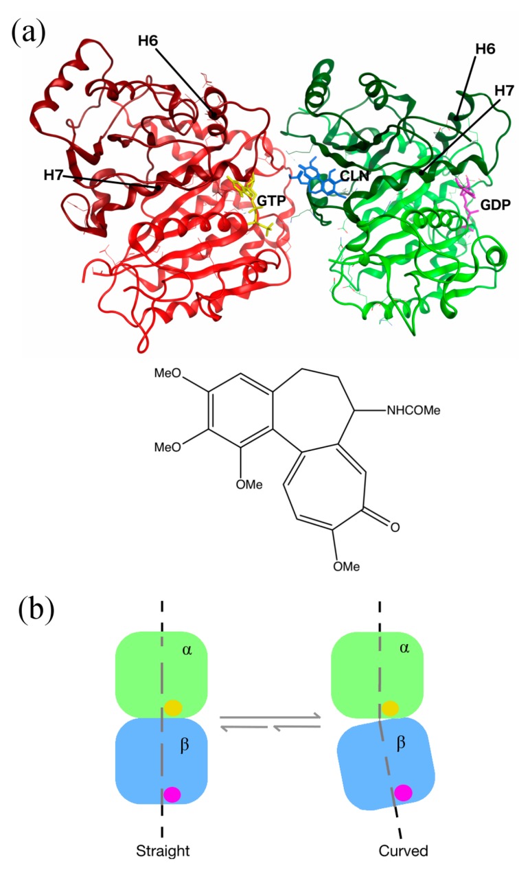
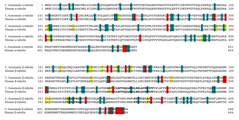
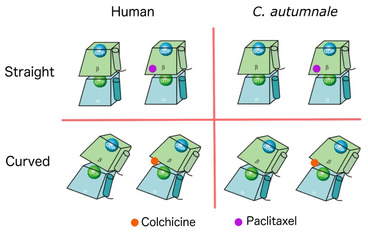
Tubulin is the target for many small-molecule natural compounds, which alter microtubules dynamics, and lead to cell cycle arrest and apoptosis. One of these compounds is colchicine, a plant alkaloid produced by Colchicum autumnale. While C. autumnale produces a potent cytotoxin, colchicine, and expresses its target protein, it is immune to colchicine's cytotoxic action and the mechanism of this resistance is hitherto unknown. In the present paper, the molecular mechanisms responsible for colchicine resistance in C. autumnale are investigated and compared to human tubulin. To this end, homology models for C. autumnale α-β tubulin heterodimer are created and molecular dynamics (MD) simulations together with molecular mechanics Poisson-Boltzmann calculations (MM/PBSA) are performed to determine colchicine's binding affinity for tubulin. Using our molecular approach, it is shown that the colchicine-binding site in C. autumnale tubulin contains a small number of amino acid substitutions compared to human tubulin. However, these substitutions induce significant reduction in the binding affinity for tubulin, and subsequently fewer conformational changes in its structure result. It is suggested that such small conformational changes are insufficient to profoundly disrupt microtubule dynamics, which explains the high resistance to colchicine by C. autumnale.

Keywords: C. autumnale; binding site; colchicine; cytotoxicity; molecular modeling; tubulin.


Conflict of interest statement

The authors declare no conflict of interest.

Figures

Figure 1
Figure 1
Structure of human tubulin heterodimer bound to colchicine, guanosine triphosphate (GTP) and guanosine diphosphate (GDP). (a) Location of the colchicine-binding site: α and β tubulins are presented in red and green ribbon structures, respectively, with the intermediate domains in darker colors. GTP and GDP are shown in yellow and purple, respectively. Colchicine (CLN) is represented in blue, with a zoom on its chemical structure on the picture below (the image was prepared with MOE2012.10 [11], adapted from the Protein Data Bank (PDB) ID:1SA0). (b) Schematic representation of the conformational changes in tubulin, undergoing from straight to curved structures. The α subunit is bound to GTP (yellow ball), and the β subunit to GDP (purple ball). The tubulin dimer representation was redrawn based on the information obtained from Ravelli et al.’s 2004 study [6].
Figure 2
Figure 2
Sequence alignment of the Colchicum autumnale tubulin with human tubulin. The C. autumnale α tubulin sequence (1KP SFCT) was aligned to the human α chain tubulin (TUBA1C) (PDB ID: Q9BQE3), and the C. autumnale β tubulin sequence (1KP: NHIX) was aligned to the human tubulin βI isoform (TUBB) (PDB ID: P07437). The sequence alignment was performed using ClustalX 2.0 [14]. Identical residues are shown in a white background. “Strongly” conserved residues within the same group are shown in cyan, and “weakly” conserved residues (within the same group, but different charge/structure) are shown in yellow. Residue substitutions are shown in red. The residues identified in the colchicine-binding domain are represented with bold letters.
Figure 3
Figure 3
Schematic representation of each of the eight homology models constructed for this study. The α tubulin is represented in blue, and the β tubulin in green. Each subunit is bound to a nucleotide. The α subunit is bound to GTP (green ball), and the β subunit to GDP (blue ball). Colchicine is represented in orange, and paclitaxel in purple. The graphical representation of the tubulin dimers was generated based on the information obtained from Krebs et al.’s 2005 study [16].
Figure 4
Figure 4
The root-mean-square deviation (RMSD) from the initial minimized structures for each of the studied systems. Plot (a) is human tubulin in straight conformation without ligand, plot (b) is human tubulin in straight conformation bound to paclitaxel, plot (c) is human tubulin in curved conformation without ligand, plot (d) is human tubulin in curved conformation bound to colchicine. Plot (e) is C. autumnale tubulin in straight conformation without ligand, plot (f) is C. autumnale tubulin in straight conformation bound to paclitaxel, plot (g) is C. autumnale tubulin in curved conformation without ligand, plot (h) is C. autumnale tubulin in curved conformation bound to colchicine. RMSD analyses are shown for both full-length protein (black curved) and the ligand-binding domain when the ligand is present (red curve).
Figure 5
Figure 5
Comparison between the holo and apo forms of tubulin. (a,b): average atomic fluctuations per residue in human (a) and in C. autumnale (b) tubulins. The tubulin heterodimer is represented in red in the presence of colchicine, and in black in the absence of colchicine. Key amino acids involved in colchicine binding are depicted on the graphs. (c,d): overlay of the conformations of the colchicine-binding domain of β tubulin in the presence and absence of colchicine. The conformations of the intermediate domain of the human β-tubulin are shown in (c) and the ones of the C. autumnale β tubulin are depicted in (d). The holo form of tubulin bound to colchicine is colored in blue, and the apo form in red.

References

    1. Hyams J.S., Lloyd C.W. Microtubules. Wiley-Liss; New York, NY, USA: 1994.
    1. Mitchison T., Kirschner M. Dynamic instability of microtubule growth. Nature. 1984;312:237–242. doi: 10.1038/312237a0. - DOI - PubMed
    1. Jordan M.A., Wilson L. Microtubules as a target for anticancer drugs. Nat. Rev. Cancer. 2004;4:253–265. doi: 10.1038/nrc1317. - DOI - PubMed
    1. Wallace S.L. Colchicine. Colchicine. 1974;3:369–381. doi: 10.1016/0049-0172(74)90006-7. - DOI - PubMed
    1. Weisenberg R.C., Borisy G.G., Taylor E.W. The colchicine-binding protein of mammalian brain and its relation to microtubules. Biochemistry. 1968;7:4466–4479. doi: 10.1021/bi00852a043. - DOI - PubMed