. 2017 Aug 2;18(8):1686.
doi: 10.3390/ijms18081686.

Intramitochondrial Ascorbic Acid Enhances the Formation of Mitochondrial Superoxide Induced by Peroxynitrite via a Ca2+-Independent Mechanism

Affiliations

Intramitochondrial Ascorbic Acid Enhances the Formation of Mitochondrial Superoxide Induced by Peroxynitrite via a Ca2+-Independent Mechanism

Andrea Guidarelli et al. Int J Mol Sci. .

Abstract

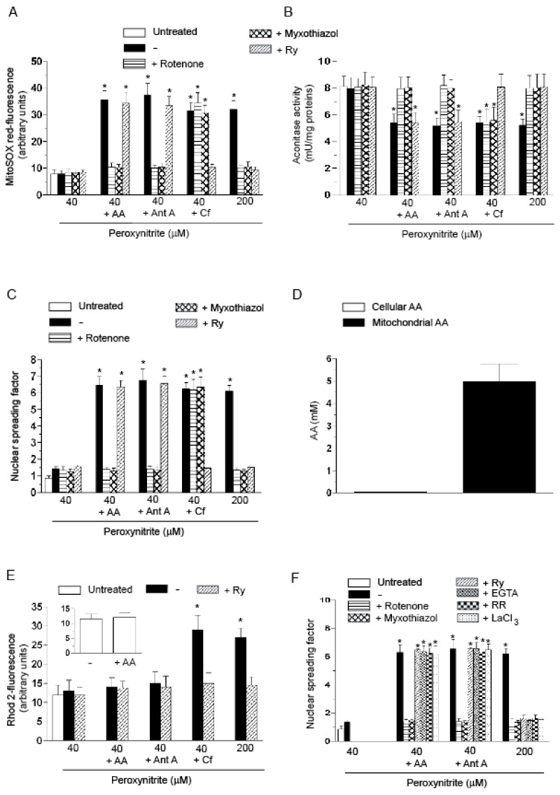
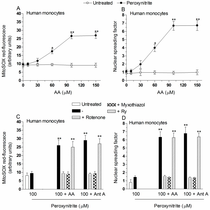
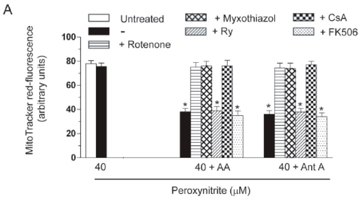
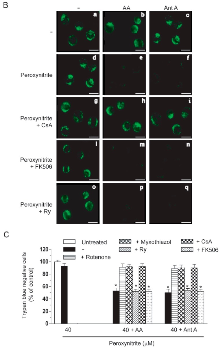
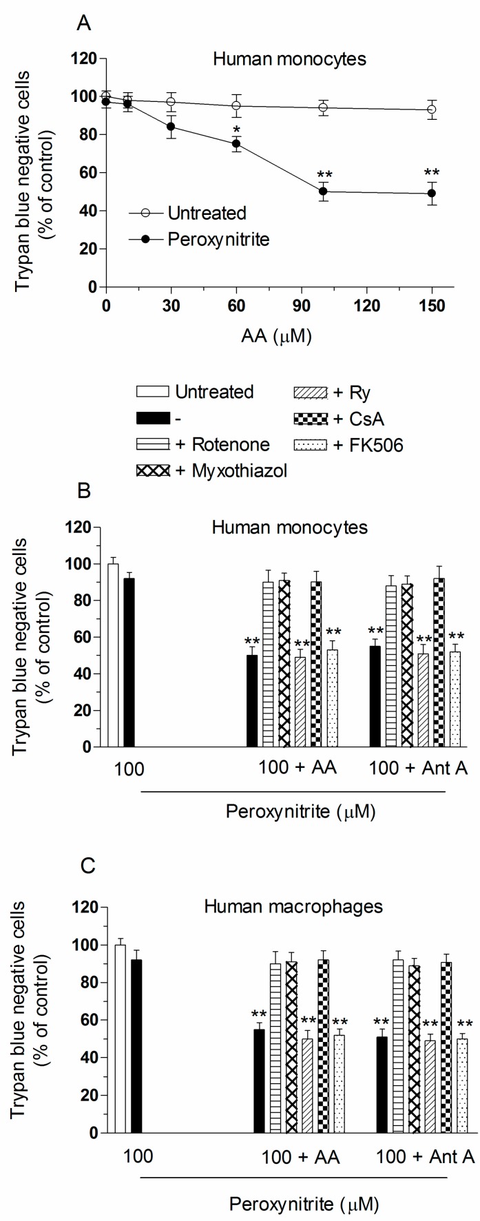
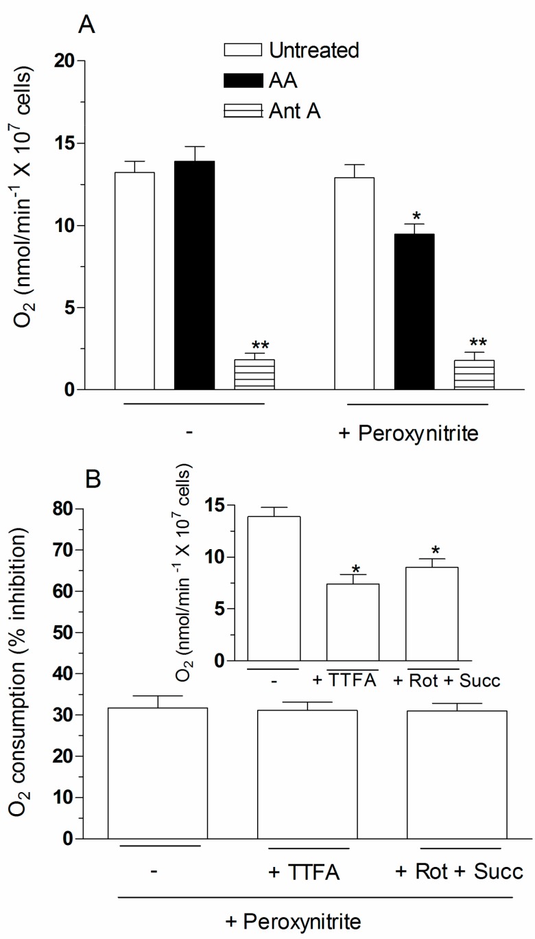
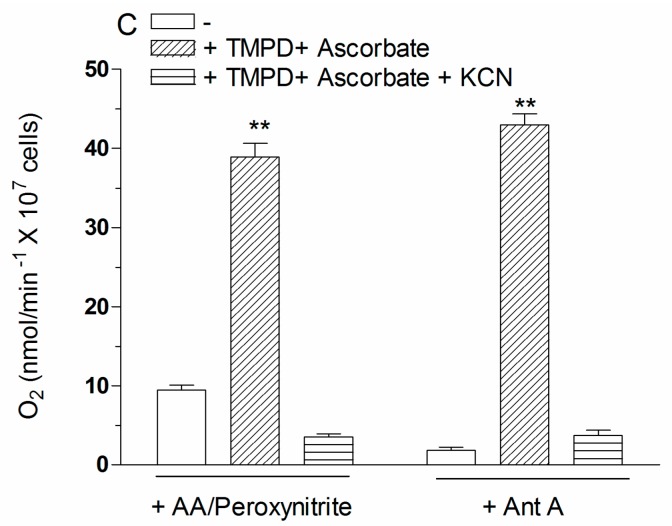
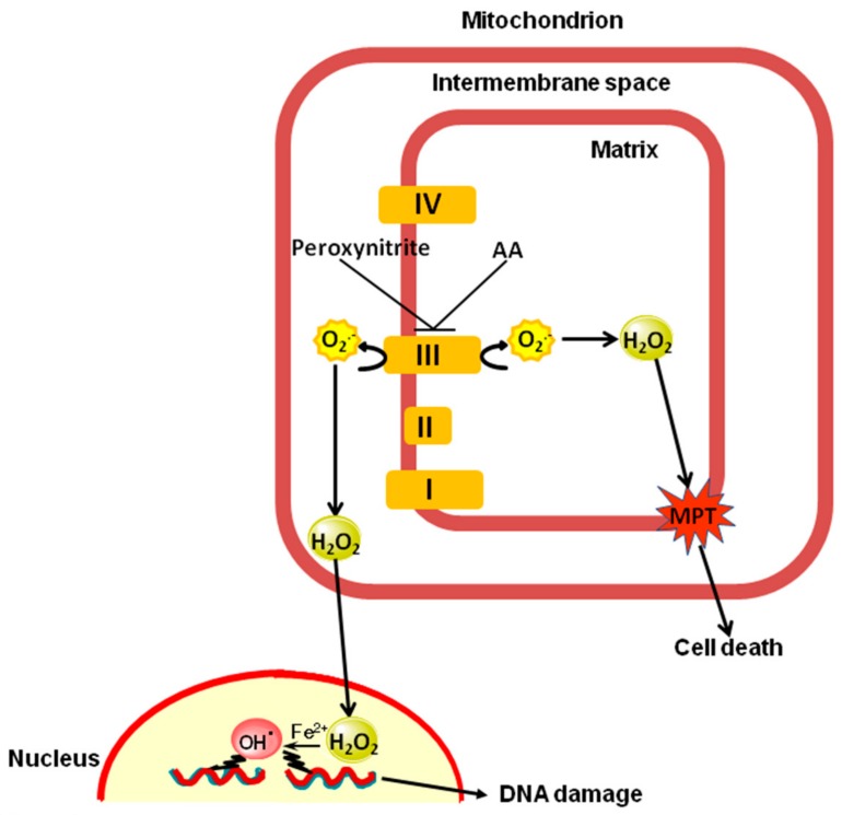
Exposure of U937 cells to peroxynitrite promotes mitochondrial superoxide formation via a mechanism dependent on both inhibition of complex III and increased mitochondrial Ca2+ accumulation. Otherwise inactive concentrations of the oxidant produced the same maximal effects in the presence of either complex III inhibitors or agents mobilizing Ca2+ from the ryanodine receptor and enforcing its mitochondrial accumulation. l-Ascorbic acid (AA) produced similar enhancing effects in terms of superoxide formation, DNA strand scission and cytotoxicity. However, AA failed to enhance the intra-mitochondrial concentration of Ca2+ and the effects observed in cells supplemented with peroxinitrite, while insensitive to manipulations preventing the mobilization of Ca2+, or the mitochondrial accumulation of the cation, were also detected in human monocytes and macrophages, which do not express the ryanodine receptor. In all these cell types, mitochondrial permeability transition-dependent toxicity was detected in cells exposed to AA/peroxynitrite and, based on the above criteria, these responses also appeared Ca2+-independent. The enhancing effects of AA are therefore similar to those mediated by bona fide complex III inhibitors, although the vitamin failed to directly inhibit complex III, and in fact enhanced its sensitivity to the inhibitory effects of peroxynitrite.

Keywords: DNA damage; ascorbic acid; complex III; mitochondrial dysfunction; mitochondrial superoxide; peroxynitrite.


Conflict of interest statement

The authors declare no conflict of interest.

Figures

Figure 1
Figure 1
The enhancing effects of AA in U937 cells exposed to low concentrations of peroxynitrite: evidence for a Ca2+-independent mechanism. (AC) The cells were treated as shown in the figure and then exposed for: 10 min (A,B); or 30 min (C) to the indicated concentrations of peroxynitrite. AA (3 µM) was given to the cells 15 min prior to peroxynitrite. Pre-exposure to antimycin A (Ant A, 1 µM), or Cf (10 mM), was instead of only 5 min. In some experiments, rotenone (0.5 µM), myxothiazol (5 µM) or Ry (ryanodine) (20 µM) were added to the cells 5 min prior addition of AA, Ant A or Cf. After treatments, the cells were analyzed for: MitoSOX red-fluorescence (A); aconitase activity (B); and DNA strand scission (C); (D) the cells were exposed for 15 min to AA and immediately analyzed for their cellular and mitochondrial content of the vitamin; (E) Rhod 2-AM (acetoxymethyl) pre-loaded cells were treated for 10 min, as indicated in the figure, and analyzed as detailed in the Materials and Methods Section. The inset shows the Rhod 2-fluorescence response observed in cells exposed for 15 min to 0 or AA; (F) cells were permeabilized with digitonin and exposed for 10 min to the indicated concentrations of peroxynitrite alone or associated with rotenone, myxothiazol, Ry, EGTA (ethylene glycol-bis(β-aminoethylether)-N,N,N′,N′-tetraacetic acid) (10 µM), RR (ruthenium red) (200 nM) or LaCl3 (100 µM). In some experiments, the cells were exposed to AA prior to permeabilization. In other experiments, the cells were supplemented with Ant A immediately after permeabilization. Results represent the means ± SD calculated from at least three separate experiments. * p < 0.001 as compared to untreated cells (one-way ANOVA followed by Dunnett’s test).
Figure 2
Figure 2
The enhancing effects of AA are also observed in human monocytes or macrophages exposed to peroxynitrite. Human monocytes were pre-exposed for 15 min to increasing concentrations of AA and then treated for a further: 10 min (A); or 30 min (B) with 100 µM peroxynitrite. After treatments, the cells were analyzed for: MitoSOX red-fluorescence (A); and DNA damage (B). Results represent the means ± SD calculated from at least three separate experiments using monocytes from three different donors. * p < 0.01 or ** p < 0.001 as compared to untreated cells (two-way ANOVA followed by Bonferroni’s test). Human monocytes (C,D); or macrophages (E,F) were pre-exposed for 15 min to 100 µM AA, or for 5 min to antimycin A (Ant A), and then treated for a further: 10 min (C,E); or 30 min (D,F) with 100 µM peroxynitrite. In some experiments, rotenone, myxothiazol, or Ry, were given to the cultures prior to peroxynitrite. After treatments, the cells were analyzed for: MitoSOX red-fluorescence (C,E); and DNA damage (D,F). Results represent the means ± SD calculated from at least three separate experiments using monocytes (or monocyte-derived macrophages) from three different donors. ** p < 0.001 as compared to untreated cells (one-way ANOVA followed by Dunnett’s test).
Figure 2
Figure 2
The enhancing effects of AA are also observed in human monocytes or macrophages exposed to peroxynitrite. Human monocytes were pre-exposed for 15 min to increasing concentrations of AA and then treated for a further: 10 min (A); or 30 min (B) with 100 µM peroxynitrite. After treatments, the cells were analyzed for: MitoSOX red-fluorescence (A); and DNA damage (B). Results represent the means ± SD calculated from at least three separate experiments using monocytes from three different donors. * p < 0.01 or ** p < 0.001 as compared to untreated cells (two-way ANOVA followed by Bonferroni’s test). Human monocytes (C,D); or macrophages (E,F) were pre-exposed for 15 min to 100 µM AA, or for 5 min to antimycin A (Ant A), and then treated for a further: 10 min (C,E); or 30 min (D,F) with 100 µM peroxynitrite. In some experiments, rotenone, myxothiazol, or Ry, were given to the cultures prior to peroxynitrite. After treatments, the cells were analyzed for: MitoSOX red-fluorescence (C,E); and DNA damage (D,F). Results represent the means ± SD calculated from at least three separate experiments using monocytes (or monocyte-derived macrophages) from three different donors. ** p < 0.001 as compared to untreated cells (one-way ANOVA followed by Dunnett’s test).
Figure 3
Figure 3
AA enhances the lethal response mediated by peroxynitrite in U937 cells via a mitochondrial permeability transition (MPT)-dependent mechanism uniquely sensitive to rotenone, myxothiazol or CsA. (A) U937 cells were pre-exposed for 15 min to AA, or for 5 min to antimycin A (Ant A), and then treated for 10 min with 40 μM peroxynitrite. In some experiments, rotenone, myxothiazol, Ry, CsA (0.5 µM) and FK506 (1 µM) were given to the cultures prior to peroxynitrite. After treatments, the cells were analyzed for MitoTracker red CMXRos-fluorescence; (B) representative micrographs of U937 cells loaded for 15 min with 1 µM calcein-acetoxymethyl ester and 1 mM CoCl2, washed and then post-incubated for a further 10 min with or without peroxynitrite, alone or associated with the additions indicated in the figure. The micrographs are representative of at least three separate experiments. Scale bars represent 20 µm; (C) cells were treated as detailed in (A) and then exposed for 60 min to peroxynitrite. After treatments, the cells were analyzed for toxicity with the trypan blue exclusion assay. Results represent the means ± SD calculated from at least three separate experiments. * p < 0.001 as compared to untreated cells (one-way ANOVA followed by Dunnett’s test).
Figure 3
Figure 3
AA enhances the lethal response mediated by peroxynitrite in U937 cells via a mitochondrial permeability transition (MPT)-dependent mechanism uniquely sensitive to rotenone, myxothiazol or CsA. (A) U937 cells were pre-exposed for 15 min to AA, or for 5 min to antimycin A (Ant A), and then treated for 10 min with 40 μM peroxynitrite. In some experiments, rotenone, myxothiazol, Ry, CsA (0.5 µM) and FK506 (1 µM) were given to the cultures prior to peroxynitrite. After treatments, the cells were analyzed for MitoTracker red CMXRos-fluorescence; (B) representative micrographs of U937 cells loaded for 15 min with 1 µM calcein-acetoxymethyl ester and 1 mM CoCl2, washed and then post-incubated for a further 10 min with or without peroxynitrite, alone or associated with the additions indicated in the figure. The micrographs are representative of at least three separate experiments. Scale bars represent 20 µm; (C) cells were treated as detailed in (A) and then exposed for 60 min to peroxynitrite. After treatments, the cells were analyzed for toxicity with the trypan blue exclusion assay. Results represent the means ± SD calculated from at least three separate experiments. * p < 0.001 as compared to untreated cells (one-way ANOVA followed by Dunnett’s test).
Figure 4
Figure 4
AA enhances the lethal response mediated by peroxynitrite in human monocytes or macrophages via a mechanism uniquely sensitive to rotenone, myxothiazol, or CsA. (A) Human monocytes were pre-exposed for 15 min to increasing concentrations of AA and then treated for a further 60 min with 0 or 100 µM peroxynitrite. After treatments, the cells were analyzed for toxicity with the trypan blue exclusion assay. Results represent the means ± SD calculated from at least three separate experiments from three different donors. * p < 0.01 or ** p < 0.001 as compared to untreated cells (two-way ANOVA followed by Bonferron’s test). Human monocytes (B); and macrophages (C) were treated as shown in the figure and then exposed for 60 min to peroxynitrite. After treatments, the cells were analyzed for toxicity with the trypan blue exclusion assay. Results represent the means ± SD calculated from at least three separate experiments using monocytes (or monocyte-derived macrophages) from three different donors. ** p < 0.001 as compared to untreated cells (one-way ANOVA followed by Dunnett’s test).
Figure 5
Figure 5
Effect of peroxynitrite on the oxygen consumption of cells preloaded with AA. (A) The cells were pre-exposed for 15 min to 0 or 3 µM AA and then analyzed for 3 min for oxygen consumption and for 3 more min after addition of peroxynitrite (80 µM). Oxygen consumption was also measured in cells supplemented with antimycin A (3 min) and then exposed to peroxynitrite (3 min). Results represent the means ± SD calculated from at least three separate experiments. * p < 0.01 or ** p < 0.001 as compared to untreated cells (one-way ANOVA followed by Dunnett’s test); (B) the cells were first pre-exposed to AA and then analysed for 3 min for oxygen consumption in the absence of other additions, or in the presence of either 250 µM TFA (trifluoroacetic acid) or 0.5 µM rotenone (Rot)/6 mM succinate (Succ) (Inset). Rotenone markedly reduced (89.13%) oxygen consumption in the absence of succinate. The extent of inhibition of oxygen consumption detected after addition of peroxynitrite (3 min) in each of these conditions was also tested (main graph). Results represent the means ± SD calculated from at least three separate experiments. * p < 0.01 as compared to untreated cells preloaded with AA (one-way ANOVA followed by Dunnett’s test); (C) the cells were pre-exposed to AA and then treated with peroxynitrite to promote inhibition of oxygen consumption, as detailed in the legend to Figure 5A. Treatment with peroxynitrite was also performed in the presence of 0.4 mM (tetramethyl-p-phenylenediamine) TMPD/1 mM ascorbate to directly activate complex IV. Sensitivity to KCN (1 mM) was then determined to link the increased oxygen consumption to stimulation of complex IV. For comparison, the effect of TMPD/ascorbate with or without KCN was also tested in untreated cells, in which oxygen consumption was suppressed by addition of antimycin A. Results represent the means ± SD calculated from at least three separate experiments. ** p < 0.001 as compared to their respective control (one-way ANOVA followed by Dunnett’s test).
Figure 5
Figure 5
Effect of peroxynitrite on the oxygen consumption of cells preloaded with AA. (A) The cells were pre-exposed for 15 min to 0 or 3 µM AA and then analyzed for 3 min for oxygen consumption and for 3 more min after addition of peroxynitrite (80 µM). Oxygen consumption was also measured in cells supplemented with antimycin A (3 min) and then exposed to peroxynitrite (3 min). Results represent the means ± SD calculated from at least three separate experiments. * p < 0.01 or ** p < 0.001 as compared to untreated cells (one-way ANOVA followed by Dunnett’s test); (B) the cells were first pre-exposed to AA and then analysed for 3 min for oxygen consumption in the absence of other additions, or in the presence of either 250 µM TFA (trifluoroacetic acid) or 0.5 µM rotenone (Rot)/6 mM succinate (Succ) (Inset). Rotenone markedly reduced (89.13%) oxygen consumption in the absence of succinate. The extent of inhibition of oxygen consumption detected after addition of peroxynitrite (3 min) in each of these conditions was also tested (main graph). Results represent the means ± SD calculated from at least three separate experiments. * p < 0.01 as compared to untreated cells preloaded with AA (one-way ANOVA followed by Dunnett’s test); (C) the cells were pre-exposed to AA and then treated with peroxynitrite to promote inhibition of oxygen consumption, as detailed in the legend to Figure 5A. Treatment with peroxynitrite was also performed in the presence of 0.4 mM (tetramethyl-p-phenylenediamine) TMPD/1 mM ascorbate to directly activate complex IV. Sensitivity to KCN (1 mM) was then determined to link the increased oxygen consumption to stimulation of complex IV. For comparison, the effect of TMPD/ascorbate with or without KCN was also tested in untreated cells, in which oxygen consumption was suppressed by addition of antimycin A. Results represent the means ± SD calculated from at least three separate experiments. ** p < 0.001 as compared to their respective control (one-way ANOVA followed by Dunnett’s test).
Figure 6
Figure 6
The enhancing effects mediated by the mitochondrial fraction of AA in cells exposed to peroxynitrite: proposed mechanism. AA increases the vulnerability of complex III to peroxynitrite via a Ca2+-independent mechanism. The extramitochondrial effects observed under these conditions are caused by superoxide released by complex III in the intermembrane space, which readily dismutates to a diffusible species, H2O2. The intramitochondrial effects are instead mediated by superoxide formation in the matrix followed by its conversion to H2O2.

Similar articles

Cited by

References

    1. Pacher P., Beckman J.S., Liaudet L. Nitric oxide and peroxynitrite in health and disease. Physiol. Rev. 2007;87:315–424. doi: 10.1152/physrev.00029.2006. - DOI - PMC - PubMed
    1. Virag L., Szabó E., Gergely P., Szabó C. Peroxynitrite-induced cytotoxicity: Mechanism and opportunities for intervention. Toxicol. Lett. 2003;140–141:113–124. doi: 10.1016/S0378-4274(02)00508-8. - DOI - PubMed
    1. Szabó C., Ohshima H. DNA damage induced by peroxynitrite: Subsequent biological effects. Nitric Oxide. 1997;1:373–385. doi: 10.1006/niox.1997.0143. - DOI - PubMed
    1. Guidarelli A., Tommasini I., Fiorani M., Cantoni O. Essential role of the mitochondrial respiratory chain in peroxynitrite-induced strand scission of genomic DNA. IUBMB Life. 2000;50:195–201. doi: 10.1080/152165400300001516. - DOI - PubMed
    1. Guidarelli A., Sciorati C., Clementi E., Cantoni O. Peroxynitrite mobilizes calcium ions from ryanodine-sensitive stores, a process associated with the mitochondrial accumulation of the cation and the enforced formation of species mediating cleavage of genomic DNA. Free Radic. Biol. Med. 2006;41:154–164. doi: 10.1016/j.freeradbiomed.2006.03.023. - DOI - PubMed

LinkOut - more resources