. 2017 Aug 2;9(8):239.
doi: 10.3390/toxins9080239.

Insights into the Mechanisms Involved in Strong Hemorrhage and Dermonecrosis Induced by Atroxlysin-Ia, a PI-Class Snake Venom Metalloproteinase

Affiliations

Insights into the Mechanisms Involved in Strong Hemorrhage and Dermonecrosis Induced by Atroxlysin-Ia, a PI-Class Snake Venom Metalloproteinase

Luciana Aparecida Freitas-de-Sousa et al. Toxins (Basel). .

Abstract

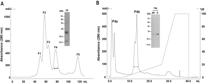
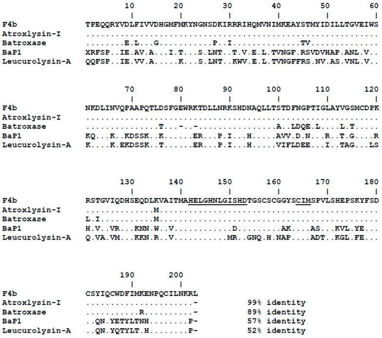
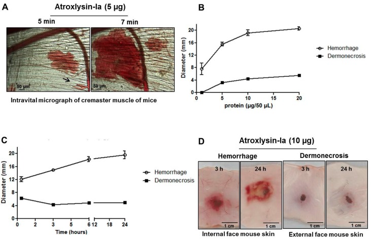
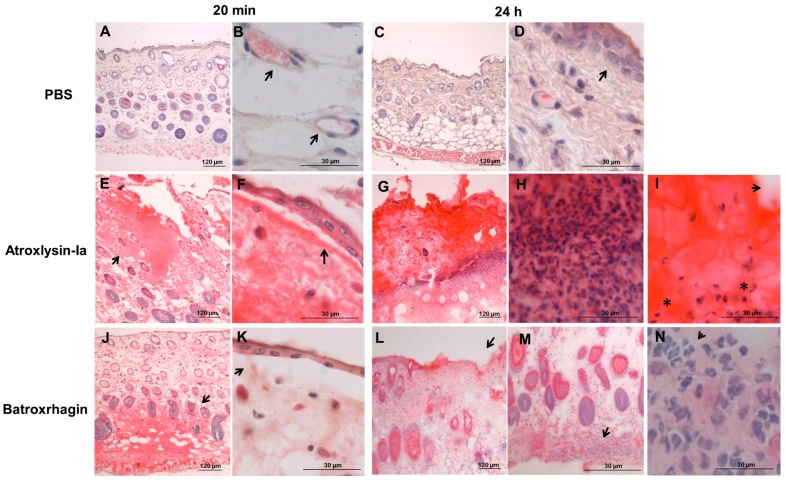
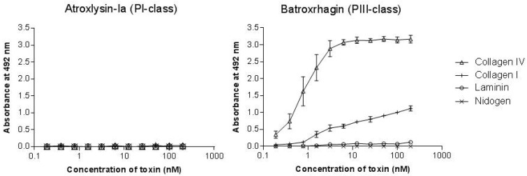
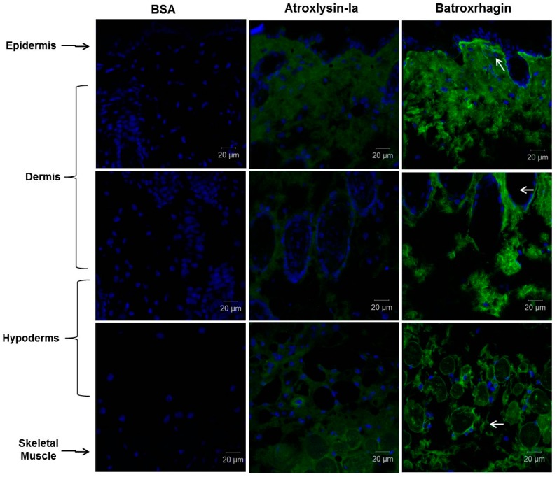
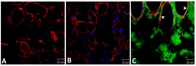
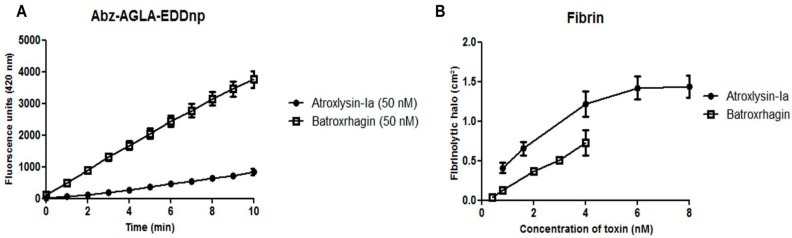
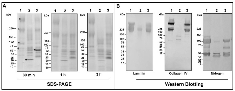
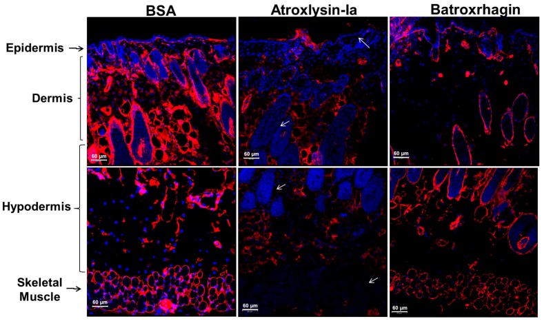
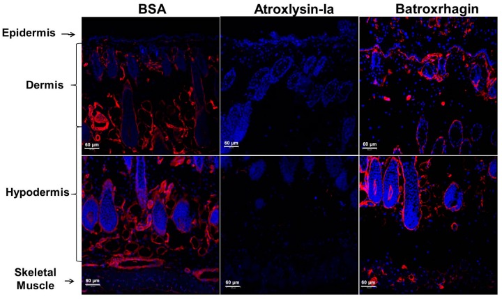
Hemorrhage is the most prominent effect of snake venom metalloproteinases (SVMPs) in human envenomation. The capillary injury is a multifactorial effect caused by hydrolysis of the components of the basement membrane (BM). The PI and PIII classes of SVMPs are abundant in viperid venoms and hydrolyze BM components. However, hemorrhage is associated mostly with PIII-class SVMPs that contain non-catalytic domains responsible for the binding of SVMPs to BM proteins, facilitating enzyme accumulation in the tissue and enhancing its catalytic efficiency. Here we report on Atroxlysin-Ia, a PI-class SVMP that induces hemorrhagic lesions in levels comparable to those induced by Batroxrhagin (PIII-class), and a unique SVMP effect characterized by the rapid onset of dermonecrotic lesions. Atroxlysin-Ia was purified from B. atrox venom, and sequence analyses indicated that it is devoid of non-catalytic domains and unable to bind to BM proteins as collagen IV and laminin in vitro or in vivo. The presence of Atroxlysin-Ia was diffuse in mice skin, and localized mainly in the epidermis with no co-localization with BM components. Nevertheless, the skin lesions induced by Atroxlysin-Ia were comparable to those induced by Batroxrhagin, with induction of leukocyte infiltrates and hemorrhagic areas soon after toxin injection. Detachment of the epidermis was more intense in skin injected with Atroxlysin-Ia. Comparing the catalytic activity of both toxins, Batroxrhagin was more active in the hydrolysis of a peptide substrate while Atroxlysin-Ia hydrolyzed more efficiently fibrin, laminin, collagen IV and nidogen. Thus, the results suggest that Atroxlysin-Ia bypasses the binding step to BM proteins, essential for hemorrhagic lesions induced by PII- and P-III class SVMPs, causing a significantly fast onset of hemorrhage and dermonecrosis, due to its higher proteolytic capacity on BM components.

Keywords: Bothrops atrox; dermonecrosis; extracellular matrix; hemorrhage; metalloproteinase; snake; venom.


Conflict of interest statement

The authors declare no conflict of interest.

Figures

Figure 1
Figure 1
Purification of the PI-class SVMP from B. atrox venom. Venom samples (50 mg) were applied to HiPrep 16/60 Sephacryl S-200 HR column and eluted at flow rate of 0.5 mL/min in 20 mM Tris/HCl buffer, pH 7.8, containing 150 mM of NaCl (A). Fractions containing ~25 kDa proteins presenting hemorrhage and dermonecrosis activities (continuous line under the peaks) were pooled, applied into a Mono Q 5/50 GL anion exchange column and eluted at flow rate of 1 mL/min with a gradient from 0 to 1 M of NaCl (B). Pooled fractions (10 µg) indicated in the graphs by continuous line under the peaks were subjected to SDS-PAGE under reducing (R) or non-reducing conditions (NR) and silver-stained. The band indicated by * was excised and submitted to MS/MS.
Figure 2
Figure 2
Sequence alignment of the isolated toxin with snake venom metalloproteinases (SVMPs) from different species of Bothrops. Amino acid sequence of the protein isolated in the fraction F4b was aligned with PI-class SVMPs: Atroxlysin-I (P85420) and Batroxase [23] from B. atrox venom, BaP1 (P83512) from B. asper venom and Leucurolysin-A (P84907) from B. leucurus using ClustalW [30]. Dots indicate identical residues to the first sequence. Functional motifs corresponding to the zinc-binding and Met-Turn are underlined.
Figure 3
Figure 3
Hemorrhagic and dermonecrotic activities of Atroxlysin-Ia. (A) Intravital micrograph of mice cremaster muscle after local administration of Atroxlysin-Ia (5 µg) evidencing the rupture of the capillary vessels indicated by arrow. (B) Size of hemorrhagic and dermonecrotic lesions induced by different doses of Atroxlysin-Ia, evaluated at 24 h after the intradermal injection. (C) The progression of the lesions evaluated at 20 min, 3, 6 and 24 h after injection of 10 µg Atroxlysin-Ia. (D) Macroscopic view of hemorrhagic (inner dorsal skin) and dermonecrosis (outer dorsal skin) activities evaluated at 3 h and 24 h after the intradermal injection of 10 µg Atroxlysin-Ia. (A,D) Figures are representative of three independent mice. (B,C) Results are expressed as mean and SEM of three independent experiments.
Figure 4
Figure 4
Action of Atroxlysin-Ia and Batroxrhagin in the skin of mice. Histological analysis (hematoxylin and eosin staining of paraffin embedded sections) of the site of injection of 10 μg of toxins. The group injected with phosphate-buffered saline (PBS) showed the normal histological pattern of skin (A,C) with preserved capillaries (arrow in B) and the undamaged epidermis (arrow in D). Tissues injected with Atroxlysin-Ia showed hemorrhage especially in the epidermis region (arrow in E) and Batroxrhagin induced hemorrhage predominantly in the hypodermis region (arrow in J). Both toxins induced epidermis detachment (arrows in F,K). Atroxlysin-Ia induced a large lesion in the epidermis and dermis (G), which evolved to the necrosis of the tissue characterized by exacerbated inflammatory infiltrate (H), deposit of fibrin clot (arrow in I) and pycnotic nuclei of the cells (* in I). Batroxrhagin induced a loss of epidermis (arrows in L) and inflammatory infiltrate especially in the hypodermis region (arrows in M,N).
Figure 5
Figure 5
Binding of Atroxlysin-Ia and Batroxrhagin to extracellular matrix components. Microtiter plates were coated with type I and IV collagens, laminin and nidogen for 18 h at 4 °C. Different dilutions of toxins were then added to the plates and the binding was detected at 492 nm after incubation with anti-Batroxrhagin serum produced in mice, followed by anti-mouse IgG peroxidase conjugates and the enzyme substrate. The results correspond to the mean and SEM of three independent experiments.
Figure 6
Figure 6
Distribution of Atroxlysin-Ia and Batroxrhagin in the skin. Cryosections were obtained at 20 min after injection of 10 µg in mice skin. Toxins and BSA (control) were labeled with Alexa488 emitting green fluorescence. Batroxrhagin accumulated in the basement membrane of epidermis, skin structures and skeletal muscle cells (arrows). Atroxlysin-Ia shows a weaker diffuse fluorescence and BSA did not show any fluorescence. The nuclei of the cells were stained in blue (4′,6-diamidino-2-phenylindole) and sections were examined with a confocal microscope.
Figure 7
Figure 7
Co-localization of Atroxlysin-Ia and Batroxrhagin with basement membrane proteins in the skin. Cryosections were obtained at 20 min after intradermal injection of 10 μg in mice skin. BSA (A), Atroxlysin-Ia (B) and Batroxrhagin (C) were labeled with Alexa488 emitting green fluorescence. The laminin was stained with rabbit polyclonal anti-laminin labelled with Tetramethyl Rhodamine Isothiocyanate (red). Batroxrhagin co-staining with laminin is represented by yellow dots (arrows in C). The nuclei of the cells were stained blue (4′,6-diamidino-2-phenylindole) and sections were examined with a confocal microscope.
Figure 8
Figure 8
Hydrolytic activity of the Atroxlysin-Ia and Batroxrhagin in vitro. (A) Hydrolysis of synthetic peptide Abz-AGLA-EDDnp was assayed by fluorimetric assays using 200 nM of the Abz-AGLA-EDDnp peptide and 50 nM of toxins at 37 °C monitored by fluorescence at λEM 420 nm and λEX 320 nm using a spectrofluorometer. (B) The fibrinolytic activity was carried out in solidified fibrin-agarose gels. Different concentrations of the toxins were applied in the gel and the hydrolysis area was measured after incubation for 18 h at 37 °C. Results are expressed as the area of lysis (cm2). The results are the mean and standard error (SEM) from three independent experiments.
Figure 9
Figure 9
Hydrolysis of the basement membrane components by Atroxlysin-Ia and Batroxrhagin in vitro. (A) Matrigel (50 µg) was incubated with 8 µM of each toxin at 37 °C and the products of hydrolysis were evaluated by SDS-PAGE under reducing conditions. (B) Matrigel (50 µg) was incubated with 10 µg of each toxin (Atroxlysin-Ia 16 µM and of Batroxrhagin 8 µM) and the products of hydrolysis of the laminin, collagen IV and nidogen were evaluated by Western Blotting using specific antibodies to each basement membrane (BM) protein. Molecular mass markers are indicated on the left. Experimental groups are: 1—Matrigel (control); 2—Atroxlysin-Ia + Matrigel; 3—Batroxrhagin + Matrigel. The most abundant components in the control correspond to laminin α1 chain and one chain of collagen IV (a), laminin β1 and γ1 chains and the second chain of collagen IV (b) and nidogen chains (c and d) and toxin bands are marked with *.
Figure 10
Figure 10
Hydrolysis of the laminin by Atroxlysin-Ia and Batroxrhagin in vivo. Cryosections were obtained at 20 min after injection of 10 µg of toxins in mice skin. Laminin is stained in red by rabbit polyclonal anti-laminin labelled with Tetramethyl Rhodamine Isothiocyanate and the nuclei of the cells was stained blue (4′,6-diamidino-2-phenylindole). The control group (BSA) show the normal distribution of laminin in the BM of the epidermis/dermis interface, around the appendages of the skin and skeletal muscle. Atroxlysin-Ia hydrolyzed more laminin in BM (arrows) than Batroxrhagin. Sections were examined with a Confocal Microscope.
Figure 11
Figure 11
Hydrolysis of the collagen IV in the skin of mice by Atroxlysin-Ia and Batroxrhagin in vivo. The cryosections were obtained at 20 min after injection of 10 µg of toxins in mice skin. Collagen is stained in red with rabbit polyclonal anti-collagen IV labelled with Tetramethyl Rhodamine Isothiocyanate and the nuclei of the cells was stained blue (4′,6-diamidino-2-phenylindole). The control group (BSA) show the normal distribution of collagen IV in the BM of the epidermis/dermis interface, around the appendages of the skin and skeletal muscle and Atroxlysin-Ia hydrolyzed more collagen IV in BM than Batroxrhagin. Sections were examined with a Confocal Microscope.

Similar articles

Cited by

References

    1. Hati R., Mitra P., Sarker S., Bhattacharyya K.K. Snake venom hemorrhagins. Crit. Rev. Toxicol. 1999;29:1–19. doi: 10.1080/10408449991349168. - DOI - PubMed
    1. Gutiérrez J.M., Romero M., Núñez J., Chaves F., Borkow G., Ovadia M. Skeletal muscle necrosis and regeneration after injection of bah1, a hemorrhagic metalloproteinase isolated from the venom of the snake Bothrops asper (terciopelo) Exp. Mol. Pathol. 1995;62:28–41. doi: 10.1006/exmp.1995.1004. - DOI - PubMed
    1. Gutiérrez J.M., Rucavado A., Chaves F., Díaz C., Escalante T. Experimental pathology of local tissue damage induced by Bothrops asper snake venom. Toxicon. 2009;54:958–975. doi: 10.1016/j.toxicon.2009.01.038. - DOI - PubMed
    1. Jiménez N., Escalante T., Gutiérrez J.M., Rucavado A. Skin pathology induced by snake venom metalloproteinase: Acute damage, revascularization, and re-epithelization in a mouse ear model. J. Investig. Dermatol. 2008;128:2421–2428. doi: 10.1038/jid.2008.118. - DOI - PubMed
    1. Moura-da-Silva A.M., Butera D., Tanjoni I. Importance of snake venom metalloproteinases in cell biology: Effects on platelets, inflammatory and endothelial cells. Curr. Pharm. Des. 2007;13:2893–2905. doi: 10.2174/138161207782023711. - DOI - PubMed

Publication types