. 2017 Aug 2;10(8):892.
doi: 10.3390/ma10080892.

Mechanical Contact Characteristics of PC3 Human Prostate Cancer Cells on Complex-Shaped Silicon Micropillars

Affiliations

Mechanical Contact Characteristics of PC3 Human Prostate Cancer Cells on Complex-Shaped Silicon Micropillars

Brandon B Seo et al. Materials (Basel). .

Abstract

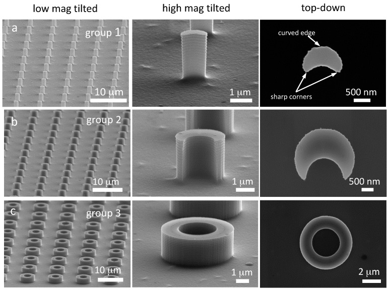
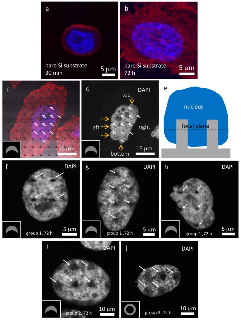
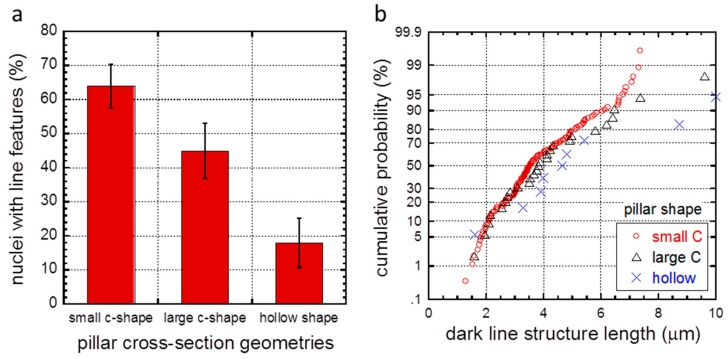
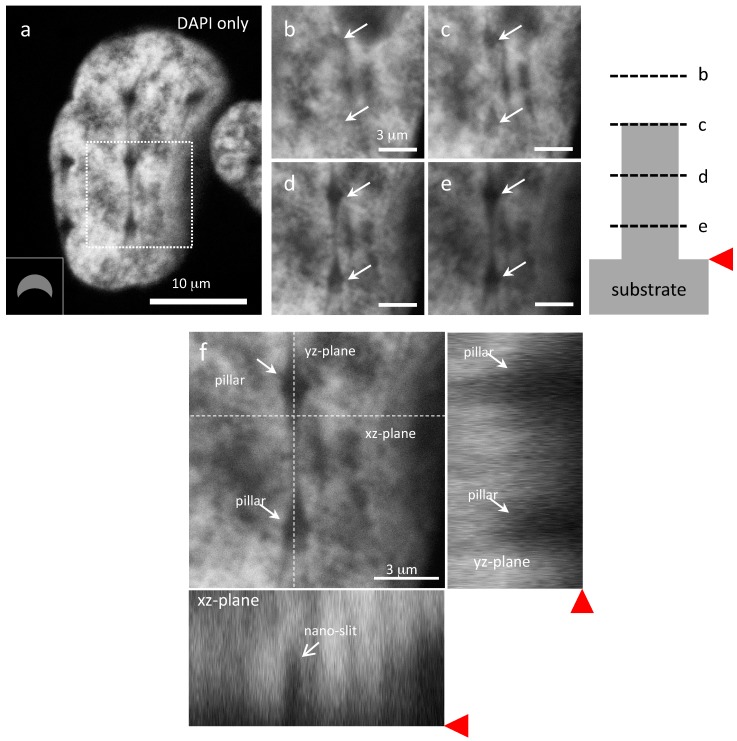
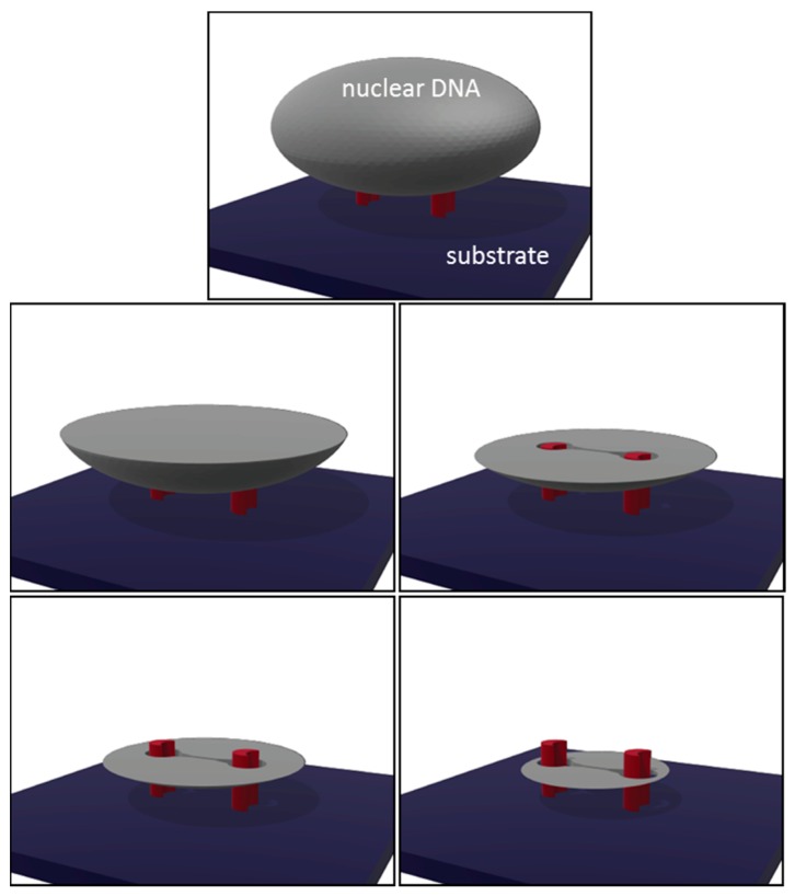
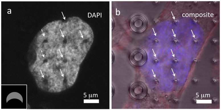
In this study we investigated the contact characteristics of human prostate cancer cells (PC3) on silicon micropillar arrays with complex shapes by using high-resolution confocal fluorescence microscopy techniques. These arrays consist of micropillars that are of various cross-sectional geometries which produce different deformation profiles in adherent cells. Fluorescence micrographs reveal that some DAPI (4',6-diamidino-2-phenylindole)-stained nuclei from cells attached to the pillars develop nanometer scale slits and contain low concentrations of DNA. The lengths of these slits, and their frequency of occurrence, were characterized for various cross-sectional geometries. These DNA-depleted features are only observed in locations below the pillar's top surfaces. Results produced in this study indicate that surface topography can induce unique nanometer scale features in the PC3 cell.

Keywords: PC3 cells; contact mechanics; deformation; nanoindentation; pillars; silicon.


Conflict of interest statement

The authors declare no conflicts of interest.

Figures

Figure 1
Figure 1
Schematic drawings of (a) spherical and (b) cylindrical tip contact geometries.
Figure 2
Figure 2
SEM micrographs of silicon pillars with complex cross-sectional geometries examined in this work: (a) group 1, small C-shaped pillars; (b) group 2, large C-shaped pillars; and (c) group 3, hollow pillars.
Figure 3
Figure 3
Confocal fluorescence micrographs of PC3 cells incubated for (a) 30 min and (b) 72 h on bare Si substrate; (c) Composite and (d) DAPI-only micrographs of PC3 cells incubated for 72 h on small C-shaped pillars (group 1). Pillar locations are indicated by solid arrows while bulged edges of the DAPI-stained materials are highlighted by dashed arrows in (c). A schematic drawing of the focal plane used in these micrographs is shown in (e). Representative micrographs of PC3 cells incubated for 72 h reveal fine line structures emanating from the small C-shaped pillars are displayed in (fh). Lines in (f) are connected with the adjacent pillars while lines in (g) and (h) are not. Confocal micrographs of 72 h incubated PC3 cells on large C-shaped and hollow-shaped pillars are displayed in (i,j), respectively. Inset drawings indicate the pillar orientations.
Figure 4
Figure 4
(a) Population of cell nuclei with slit features for three different pillar cross-section geometries. Slit length distributions on various shaped pillars are displayed in (b).
Figure 5
Figure 5
(a) DAPI only micrograph revealing fine line structures in cells incubated on small C-shaped pillars. High magnification images of a representative nucleus surrounding two small C-shaped pillars imaged in various focal planes are shown in (be). The corresponding focal plane locations are illustrated in the schematic drawing. Orthogonal views of this nucleus are shown in (f) revealing two pillars and a nano-slit. Red triangle pointers indicate substrate surface locations.
Figure 6
Figure 6
Schematic drawings of nuclear DNA elements (gray) surrounding two C-shaped pillars (red) at different cross-sectional planes above and below the pillars. Note the DNA materials are not in direct contact with the pillars. The tear drop-shaped openings in between the pillars indicate the slit location.
Figure 7
Figure 7
(a) DAPI only and (b) composite top-down micrographs of a cell adhered on small C-shaped and hollow pillars. The locations of the pillars are highlighted with arrows. Cells were incubated on the pattern substrate for 72 h. Inset drawing indicates the pillar orientation.

References

    1. Bettinger C.J., Langer R., Borenstein J.T. Engineering Substrate Micro- and Nanotopography to Control Cell Function. Angew. Chem. Int. Ed. Engl. 2009;48:5406–5415. doi: 10.1002/anie.200805179. - DOI - PMC - PubMed
    1. Bettinger C.J., Orrick B., Misra A., Langer R., Borenstein J.T. Microfabrication of poly (glycerol-sebacate) for contact guidance applications. Biomaterials. 2006;27:2558–2565. doi: 10.1016/j.biomaterials.2005.11.029. - DOI - PubMed
    1. Bucaro M.A., Vasquez Y., Hatton B.D., Aizenberg J. Fine-tuning the degree of stem cell polarization and alignment on ordered arrays of high-aspect-ratio nanopillars. ACS Nano. 2012;6:6222–6230. doi: 10.1021/nn301654e. - DOI - PubMed
    1. Hanson L., Zhao W., Lou H.-Y., Lin Z.C., Lee S.W., Chowdary P., Cui Y., Cui B. Vertical nanopillars for in situ probing of nuclear mechanics in adherent cells. Nat. Nanotechnol. 2015;10:554–562. - PMC - PubMed
    1. Hu W., Crouch A.S., Miller D., Aryal M., Luebke K.J. Inhibited cell spreading on polystyrene nanopillars fabricated by nanoimprinting and in situ elongation. Nanotechnology. 2010;21:385301. doi: 10.1088/0957-4484/21/38/385301. - DOI - PubMed

LinkOut - more resources