Review
. 2017 Aug 2;18(8):1683.
doi: 10.3390/ijms18081683.

Chemical Modifications of Nucleic Acid Aptamers for Therapeutic Purposes

Affiliations
Review

Chemical Modifications of Nucleic Acid Aptamers for Therapeutic Purposes

Shuaijian Ni et al. Int J Mol Sci. .

Abstract

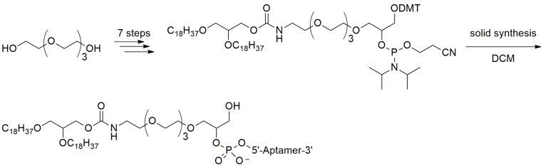
Nucleic acid aptamers have minimal immunogenicity, high chemical synthesis production, low cost and high chemical stability when compared with antibodies. However, the susceptibility to nuclease degradation, rapid excretion through renal filtration and insufficient binding affinity hindered their development as drug candidates for therapeutic applications. In this review, we will discuss methods to conquer these challenges and highlight recent developments of chemical modifications and technological advances that may enable early aptamers to be translated into clinical therapeutics.

Keywords: binding affinity; chemical modification; nuclease degradation; nucleic acid aptamer; rapid excretion.


Conflict of interest statement

The authors declare no conflict of interest.

Figures

Figure 1
Figure 1
The common strategies in the chemical modifications of nucleic acid aptamers and their purposes. Among the modifications, such as modifications on the terminals of nucleic acids, modifications on the phosphodiester linkage, modifications on the sugar ring and modifications on the bases, the 3′ end capping with inverted thymidine [6,12] and PEGylation [13] have been the common strategies in the chemical modifications of nucleic acid aptamers for development clinical therapeutics [14,15,16,17].
Figure 2
Figure 2
Four-step phosphoramidite oligodeoxynucleotide synthesis cycle (adapted from [18]). The phosphoramidite method, pioneered by Marvin Caruthers in the early 1980s, and enhanced by the application of solid-phase technology and automation, is now firmly established as the method of choice. Phosphoramidite oligonucleotide synthesis proceeds in the 3′ to 5′ direction (opposite to the 5′ to 3′ direction of DNA biosynthesis in DNA replication). One nucleotide is added per synthesis cycle. The phosphoramidite DNA synthesis cycle consists of a series of steps outlined in the figure.
Figure 3
Figure 3
Solid-phase RNA synthesis via the phosphoramidite method (adapted from [19]). In RNA synthesis, the 2′-hydroxy group is protected with TBDMS (t-butyldimethylsilyl) group, which can be removed by treatment with fluoride ion.
Figure 4
Figure 4
Solid-phase synthesis of 3′-inverted dT modified aptamers. Synthesis of 3′-inverted dT modified aptamers needs modified CPG with the 5′-hydroxyl of the first nucleoside attached, followed by chain elongation in standard 3′→5′ fashion.
Figure 5
Figure 5
Structure of the 3′-biotin conjugate. 3′-Biotin could inhibit the activity of 3′-exonuclease, which was similar to 3′-inverted dT modification. In addition, the 3′-biotin conjugates slowed down the clearance rate in blood circulation system in vivo.
Figure 6
Figure 6
2′-substitutions utilized to enhance the stability of aptamers in vivo (adapted from [39]). 2′-Substitutions can easily be incorporated into aptamers during chemical synthesis and include: (i) 2′-H; (ii) 2′-OH; (iii) 2′-NH2; (iv) 2′-F; and (v) 2′-OMe.
Figure 7
Figure 7
Structures of Locked nucleic acid (LNA), unlocked nucleic acid (UNA) and 2′-deoxy-2′-fluoro-d-arabinonucleic acid (2′-F ANA). LNA is an analog of ribonucleotide with a methylene linkage between 2′-O and 4′-C of the sugar ring. UNA misses a bond between C2′ and C3′ of the sugar ring. 2′-F ANA adopts anti-conformation with 2′-F-G.
Figure 8
Figure 8
Structures of methylphosphonate and phosphorothioate.
Figure 9
Figure 9
Fragments of oligonucleotide analogs with different types of triazole internucleotide modifications (adapted from [64]). A, B, C represent three different types of triazole internucleotide modifications.
Figure 10
Figure 10
Synthesis of the triazole internucleoside linked oligonucleotide analogs with increased resistance to DNAses and polymerases (adapted from [64]).
Figure 11
Figure 11
Structures of l-deoxyoligonucleotide (l-DNA). Mirror image aptamers are composed of non-natural l-ribose nucleotides. The molecules are initially selected from natural d-ribose aptamer libraries against a non-natural target, for example a d-peptide. Once optimized as a d-aptamer, the mirror image l-aptamer (Spiegelmer) is synthesized chemically and intrinsically bound to the natural l-target, such as a naturally occurring protein.
Figure 12
Figure 12
Structures of cholesterol-oligonucleotide conjugates (adapted from [71]). Cholesterol can be derivatized to the 5′-end of an aptamer to form a cholesterol-oligonucleotide (cholODN) conjugate. The half-time of the resulting cholODN in plasma was considerably longer than the control ODN.
Figure 13
Figure 13
Synthesis of the dialkylglycerol (DAG) modified VEGF aptamer (adapted from [72]). Liposome-anchored aptamer maintained the high binding affinity to VEGF. Moreover, the plasma residence time was considerably improved when compared with that of the original aptamer.
Figure 14
Figure 14
Addition of the aminolinker to 5′-end of the oligonucleotide and PEGylation of amino-modified oligonucleotide with 40 kDa Y-shaped PEG (n = ~450) (adapted from [74]). Amino-modified oligonucleotide could be reacted with NHS-ester-activated PEG to form oligonucleotide-PEG conjugate. Conjugation of aptamers with high molecular weight PEG could limit the rate of filtration and extended half-life up to 24–48 h.
Figure 15
Figure 15
Reaction scheme of aptamer conjugating to a 40 kDa polyethylene glycol (PEG) at the 5′-termini (adapted from [76]).
Figure 16
Figure 16
Structure of 5-BzdU (5-(N-benzylcarboxyamide)-2′-deoxyuridine).
Figure 17
Figure 17
Structures of naphtyl, triptamino and isobutyl.
Figure 18
Figure 18
Schematic of the PS2-walk library of sequence variants each containing a single PS2 modification. Modification hot spots along the phosphate backbone of the aptamer could be identified by phosphorodithioate (PS2) substitution on a single nucleotide of nucleic acid sequences.
Figure 19
Figure 19
Summary of the chemical modifications of nucleic acid aptamers.

Similar articles

Cited by

References

    1. Tuerk C., Gold L. Systematic evolution of ligands by exponential enrichment: RNA ligands to bacteriophage T4 DNA polymerase. Science. 1990;249:505–510. doi: 10.1126/science.2200121. - DOI - PubMed
    1. Ellington A.D., Szostak J.W. In vitro selection of RNA molecules that bind specific ligands. Nature. 1990;346:818. doi: 10.1038/346818a0. - DOI - PubMed
    1. Zhou J., Rossi J. Aptamers as targeted therapeutics: Current potential and challenges. Nat. Rev. Drug Discov. 2017;16:181–202. doi: 10.1038/nrd.2016.199. - DOI - PMC - PubMed
    1. Gelinas A.D., Davies D.R., Janjic N. Embracing proteins: Structural themes in aptamer-protein complexes. Curr. Opin. Struct. Biol. 2016;36:122–132. doi: 10.1016/j.sbi.2016.01.009. - DOI - PubMed
    1. Ashrafuzzaman M. Aptamers as both drugs and drug-carriers. BioMed Res. Int. 2014;2014 doi: 10.1155/2014/697923. - DOI - PMC - PubMed

MeSH terms

Substances