

The comparison of measurement between ultrasound and computed tomography for abnormal degenerative facet joints: A STROBE-compliant article
- PMID: 28767595
- PMCID: PMC5626149
- DOI: 10.1097/MD.0000000000007680
The comparison of measurement between ultrasound and computed tomography for abnormal degenerative facet joints: A STROBE-compliant article
Abstract
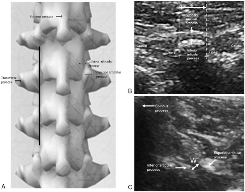
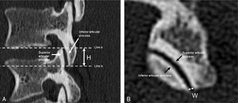
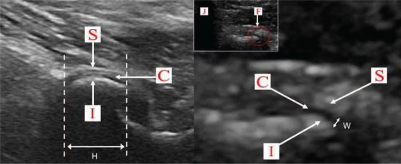
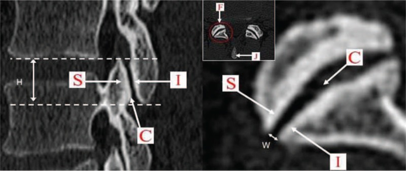
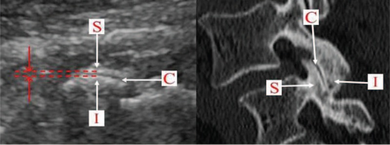
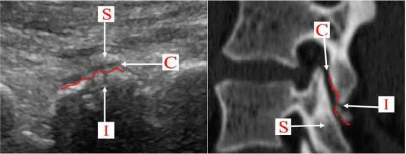
Besides the study on examining facet joints of lumbar spine by ultrasound in normal population, there has not been any related report about examining normal facet joints of lumbar spine by ultrasound so far. This study was aimed to explore the feasibility of ultrasound assessment of lumber spine facet joints by comparing ultrasound measure values of normal and degenerative lumber spine facet joints, and by comparing measure values of ultrasound and computed tomography (CT) of degenerative lumber spine facet joints.This study included 15 patients who had chronic low back pain because of degenerative change in lumbar vertebrae, and 19 volunteers who did not have low back pain or pain in the lower limb. The ultrasound measure values (height [H] and width [W]) of normal and degenerative lumber spine facet joints were compared. And the differentiation between measure values (H and W) of ultrasound and CT of degenerative lumber spine facet joints was also analyzed.The ultrasound clearly showed abnormal facet joints lesion, which was characterized by hyperostosis on the edge of joints, bone destruction under joints, and thinner or thicker articular cartilage. There were significant differences between the ultrasound measure values of the normal (H: 1.26 ± 0.03 cm, W: 0.18 ± 0.01 cm) and abnormal facet joints (H: 1.43 ± 0.05 cm, W: 0.15 ± 0.02 cm) (all P < .05). However, there were no significant differences between the measure values of the ultrasound (H: 1.43 ± 0.17 cm, W: 0.15 ± 0.03 cm) and CT (H: 1.42 ± 0.16, W: 0.14 ± 0.03) of the degenerative lumber spine facet joints (all P > .05).Ultrasound can clearly show the structure of facet joints of lumbar spine. It is precise and feasible to assess facet joints of lumbar spine by ultrasound. This study has important significance for the diagnosis of lumbar facet joint degeneration.
Conflict of interest statement
The authors report no conflicts of interest.
Figures
Similar articles
-
Micro-computed tomography, scanning electron microscopy and energy X-ray spectroscopy studies of facet joint degeneration: A comparison to clinical imaging.Micron. 2017 Sep;100:50-59. doi: 10.1016/j.micron.2017.04.011. Epub 2017 May 1. Micron. 2017. PMID: 28500930
-
Quantitative ultrasound assessment of the facet joint in the lumbar spine: a feasibility study.Ultrasound Med Biol. 2015 May;41(5):1226-32. doi: 10.1016/j.ultrasmedbio.2014.12.025. Epub 2015 Jan 28. Ultrasound Med Biol. 2015. PMID: 25638321
-
[Feasibility and accuracy of ultrasound-guided methodology in the examination of lumbar spine facet joints].Sichuan Da Xue Xue Bao Yi Xue Ban. 2013 Mar;44(2):300-2. Sichuan Da Xue Xue Bao Yi Xue Ban. 2013. PMID: 23745277 Chinese.
-
Facet Tropism in Lumbar Spine and Cervical Spine: A Systematic Review and Meta-Analysis.World Neurosurg. 2021 Mar;147:47-65. doi: 10.1016/j.wneu.2020.11.171. Epub 2020 Dec 9. World Neurosurg. 2021. PMID: 33309642
-
Does osteoarthritis of the lumbar spine cause chronic low back pain?Curr Rheumatol Rep. 2004 Feb;6(1):14-9. doi: 10.1007/s11926-004-0079-z. Curr Rheumatol Rep. 2004. PMID: 14713398 Review.
Cited by
-
Facet joint distance measurement using digital tomosynthesis while standing.J Biomech. 2025 Apr;183:112596. doi: 10.1016/j.jbiomech.2025.112596. Epub 2025 Feb 21. J Biomech. 2025. PMID: 40023053
-
Enhancing Clinical Insights: New Radiographic Grading for Lumbar Facet Joint Degeneration, A Reliability Study.JOR Spine. 2025 Jan 7;8(1):e70035. doi: 10.1002/jsp2.70035. eCollection 2025 Mar. JOR Spine. 2025. PMID: 39781088 Free PMC article.
-
Consensus Guidelines from the American Society of Pain and Neuroscience for the Use of 60-Day Peripheral Nerve Stimulation Therapy. A NEURON Living Guideline Project.J Pain Res. 2025 Jun 24;18:3117-3139. doi: 10.2147/JPR.S521788. eCollection 2025. J Pain Res. 2025. PMID: 40590046 Free PMC article.
-
Functional MRI for evaluation of hyaline cartilage extracelullar matrix, a physiopathological-based approach.Br J Radiol. 2019 Nov;92(1103):20190443. doi: 10.1259/bjr.20190443. Epub 2019 Aug 23. Br J Radiol. 2019. PMID: 31433668 Free PMC article. Review.
References
-
- Kalichman L, Hunter DJ. Lumbar facet joint osteoarthritis: a review. Semin Arthritis Rheum 2007;37:69–80. - PubMed
-
- Eubanks JD, Lee MJ, Cassinelli E, et al. Prevalence of lumbar facet arthrosis and its relationship to age, sex, and race: an anatomic study of cadaveric specimens. Spine 2007;32:2058–62. - PubMed
-
- Manchikanti L, Singh V. Review of chronic low back pain of facet joint origin. Pain Physician 2002;5:83–101. - PubMed
Publication types
MeSH terms
LinkOut - more resources
Full Text Sources
Other Literature Sources
Medical
