

Multicentric Reticulohistiocytosis with Dermatomyositis-like Eruptions
- PMID: 28768982
- PMCID: PMC5577088
- DOI: 10.2169/internalmedicine.56.8297
Multicentric Reticulohistiocytosis with Dermatomyositis-like Eruptions
Abstract
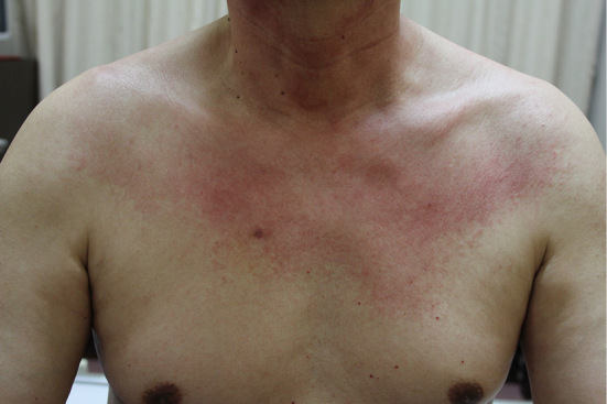
A 68-year-old man presented with polyarthritis, proximal muscle weakness, and erythema of the face, arms, neck, and anterior chest that resembled the V-neck sign. Initially, dermatomyositis (DM) was considered because of the erythema, polyarthritis, and muscle weakness. He also had mediastinal and hilar lymphadenopathy on contrast-enhanced computed tomography. Unexpectedly, a biopsy of the forehead skin revealed numerous multinucleated giant cells. A biopsy of a solitary nodule on the dorsum of his right middle finger revealed similar multinucleated giant cells with ground-glass cytoplasm, leading to the diagnosis of multicentric reticulohistiocytosis (MRH). Although MRH is rare, it should be remembered that MRH can mimic DM.
Keywords: dermatomyositis; multicentric reticulohistiocytosis; rheumatoid arthritis; sarcoidosis.
Figures


References
-
- Tajirian AL, Malik MK, Robinson-Bostom L, Lally EV. Multicentric reticulohistiocytosis. Clin Dermatol 24: 486-492, 2006. - PubMed
-
- Trotta F, Colina M. Multicentric reticulohistiocytosis and fibroblastic rheumatism. Best Pract Res Clin Rheumatol 26: 543-557, 2012. - PubMed
-
- Campbell DA, Edwards NL. Multicentric reticulohistiocytosis: systemic macrophage disorder. Baillieres Clin Rheumatol 5: 301-319, 1991. - PubMed
-
- Tait TJ, Bird HA, Ford GP. Multicentric reticulohistiocytosis: presentation with the cutaneous features of dermatomyositis. Br J Rheumatol 33: 100-101, 1994. - PubMed
Publication types
MeSH terms
LinkOut - more resources
Full Text Sources
Other Literature Sources