. 2017 Aug 2;7(1):7143.
doi: 10.1038/s41598-017-07101-2.

Characterisation of lung macrophage subpopulations in COPD patients and controls

Affiliations

Characterisation of lung macrophage subpopulations in COPD patients and controls

Jennifer A Dewhurst et al. Sci Rep. .

Abstract

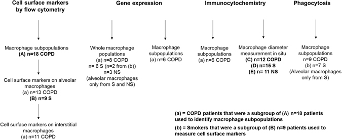
Lung macrophage subpopulations have been identified based on size. We investigated characteristics of small and large macrophages in the alveolar spaces and lung interstitium of COPD patients and controls. Alveolar and interstitial cells were isolated from lung resection tissue from 88 patients. Macrophage subpopulation cell-surface expression of immunological markers and phagocytic ability were assessed by flow cytometry. Inflammatory related gene expression was measured. Alveolar and interstitial macrophages had subpopulations of small and large macrophages based on size and granularity. Alveolar macrophages had similar numbers of small and large cells; interstitial macrophages were mainly small. Small macrophages expressed significantly higher cell surface HLA-DR, CD14, CD38 and CD36 and lower CD206 compared to large macrophages. Large alveolar macrophages showed lower marker expression in COPD current compared to ex-smokers. Small interstitial macrophages had the highest pro-inflammatory gene expression levels, while large alveolar macrophages had the lowest. Small alveolar macrophages had the highest phagocytic ability. Small alveolar macrophage CD206 expression was lower in COPD patients compared to smokers. COPD lung macrophages include distinct subpopulations; Small interstitial and small alveolar macrophages with more pro-inflammatory and phagocytic function respectively, and large alveolar macrophages with low pro-inflammatory and phagocytic ability.


Conflict of interest statement

D. Singh has received sponsorship to attend international meetings, honoraria for lecturing or attending advisory boards and research grants from various pharmaceutical companies including Almirall, AstraZeneca, Boehringer Ingelheim, Chiesi, Genentech, GlaxoSmithKline, Glenmark, Johnson and Johnson, Merck, NAPP, Novartis, Pfizer, Skypharma, Takeda, Teva, Therevance and Verona.E. Hardaker was employed by Novartis during the undertaking of this project. S. Lea, J. Dewhurst, J. Dungwa and A. Ravi declare that they have no competing interests

Figures

Figure 1
Figure 1
Flow chart describing subsets of patients. a = cells from the same COPD patients used for identifying macrophage subpopulations were used for these experiments, b = cells from the same smoking controls used for measuring cell surface markers on alveolar macrophages were used for these experiments. S: smoking controls, NS: never smokers.
Figure 2
Figure 2
Identifying macrophage subpopulations in alveolar and interstitial macrophages. Alveolar macrophage (A,B and C) and interstitial macrophage (D,E and F) subpopulations were compared and data is shown of enriched macrophages from an ex smoking COPD patient. Flow cytometry pseudocolor plots (A and D and density plots (B and E) indicating size (FSC) and granularity (SSC) were used to identify small (a) and large (b) macrophage subpopulations. H&E stained cytospins (C and F) were used to confirm the presence of small (a) and large (b) macrophages. The number of small and large macrophages present was expressed as a percentage of the total macrophage population (G). Data represents mean (SEM) of n = 18 COPD patients. Paired t test (two tailed) was performed. ***The percentage of large interstitial macrophages is significantly decreased compared to the percentage of large alveolar macrophages (p < 0.001).
Figure 3
Figure 3
The expression of macrophage markers in small macrophages compared to large macrophages. Flow cytometric analysis of enriched alveolar (A and B) and interstitial (C and D) macrophages for n = 13 and n = 11 COPD patients respectively per marker. Markers analysed were HLA-DR, CD14, CD38, CD36, CD206 and CD163. Data is expressed as the percentage of cells within each subpopulation expressing a specific marker (A and C) and the median fluorescence intensity (MFI) of each marker (B and D) and represents mean (SEM). Paired t test (two tailed) was performed for each marker. *,**,***Significantly increased between size populations within tissue compartment (p < 0.05, 0.01 and 0.001 respectively). #,##,###Significantly increased between tissue compartments for each size population (p < 0.05, 0.01 and 0.001 respectively)
Figure 4
Figure 4
The expression of macrophage markers in the large alveolar macrophage population in COPD (including smoking status). Flow cytometric analysis of large alveolar macrophages for n = 7 COPD smokers (COPDS) and n = 6 COPD ex-smokers (COPDE). Markers analysed were HLA-DR (A), CD206 (B), CD163 (C), CD14 (D), CD38 (E) and CD36 (F). Data is expressed as the median fluorescence intensity (MFI) of each marker and represents mean (SEM). Unpaired t test (Two-tailed) was performed for each marker. ***Significantly different compared to expression of the same marker on large alveolar macrophages from COPDE patients (p < 0.001).
Figure 5
Figure 5
The diameter of alveolar and interstitial macrophages in lung resected tissue. Alveolar (AM) and interstitial (IM) macrophages from never smokers (NS) (n = 11) (A), smoking controls (S) (n = 15) (B) and COPD patients (COPD) (n = 12) (C) were identified in FFPE resected lung tissue by co-expression of CX3CR1 and CD14 and the diameter of each cell was measured. Data are shown as the percentage of macrophage population measuring a certain diameter (A-C) or individual cell diameters for IMs (D) and AMs (E) with median and interquartile range. Mann-whitney t-tests were performed between IM and AMs. One way ANOVA followed by Tukey-Kramer Multiple Comparisons Test were performed between subject groups. **,***Significant difference (p < 0.01, < 0.001 respectively)
Figure 6
Figure 6
Expression of CXCL1 and TLR3 protein and phagocytosis of pHrodo E. coli BioParticles in macrophage subpopulations. Alveolar (AM) and interstitial (IM) macrophages were isolated using EasySep monocyte enrichment kit. Cells were used to generate cytospin slides (A and B) or exposed to pHrodo E. coli BioParticles for 1 h in a shaking incubator (C and D). Percentage of small alveolar, large alveolar and small interstitial macrophages expressing CXCL1 protein and TLR3 protein are shown in (A and B) respectively. Data represents mean (SEM) of n = 6 COPD patients. Phagocytosis was analysed by flow cytometry and is represented by (C): % of pHrodo bright macrophages within each subpopulation and (D): median fluorescence intensity (MFI) of each subpopulation relative to the negative control. Data are shown as mean (SEM) of n = 9 COPD patients One way ANOVA followed by Tukey-Kramer Multiple Comparisons Test was performed. *,**,***Significantly different compared to expression of the same protein on large alveolar macrophages (p < 0.05, < 0.01 and <0.001 respectively). #,##Significantly different compared to the percentage of pHrodo bright macrophages or MFI of other macrophage subpopulations (p < 0.05, <0.01 respectively).

References

    1. Vogelmeier CF, et al. Global Strategy for the Diagnosis, Management, and Prevention of Chronic Obstructive Lung Disease 2017 Report. GOLD Executive Summary. Am J Respir Crit Care Med. 2017;195:557–582. doi: 10.1164/rccm.201701-0218PP. - DOI - PubMed
    1. Hogg JC, et al. The nature of small-airway obstruction in chronic obstructive pulmonary disease. N Engl J Med. 2004;350:2645–2653. doi: 10.1056/NEJMoa032158. - DOI - PubMed
    1. Di Stefano A, et al. Severity of airflow limitation is associated with severity of airway inflammation in smokers. Am J Respir Crit Care Med. 1998;158:1277–1285. doi: 10.1164/ajrccm.158.4.9802078. - DOI - PubMed
    1. Fels AO, Cohn ZA. The alveolar macrophage. J Appl Physiol. 1986;60:353–369. - PubMed
    1. Hoppstadter, J. et al. Differential cell reaction upon Toll-like receptor 4 and 9 activation in human alveolar and lung interstitial macrophages. Respir Res 11, doi:10.1186/1465-9921-11-124 (2010). - PMC - PubMed

Publication types

MeSH terms