

Cavernous hemangioma of the orbit: an unusual acute presentation
- PMID: 28769595
- PMCID: PMC5533564
- DOI: 10.2147/IMCRJ.S133284
Cavernous hemangioma of the orbit: an unusual acute presentation
Abstract
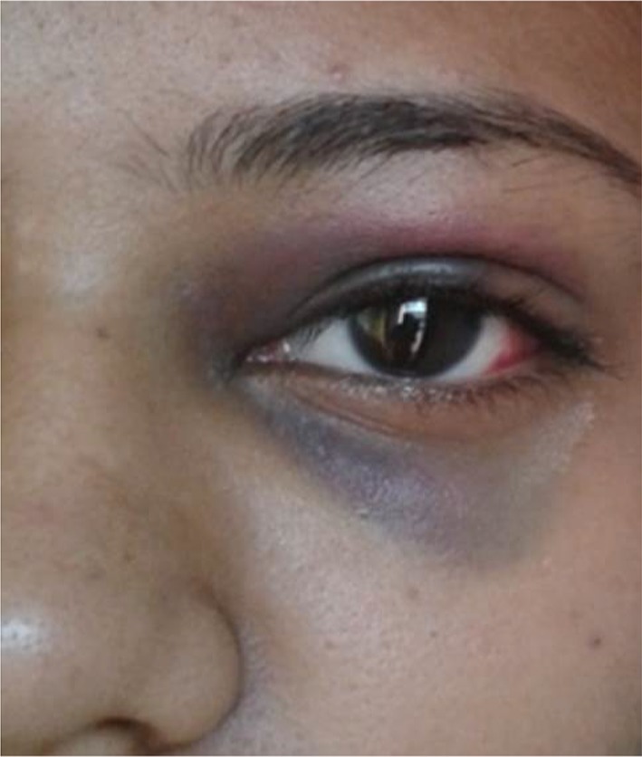
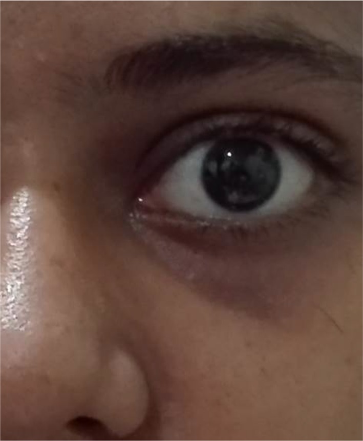
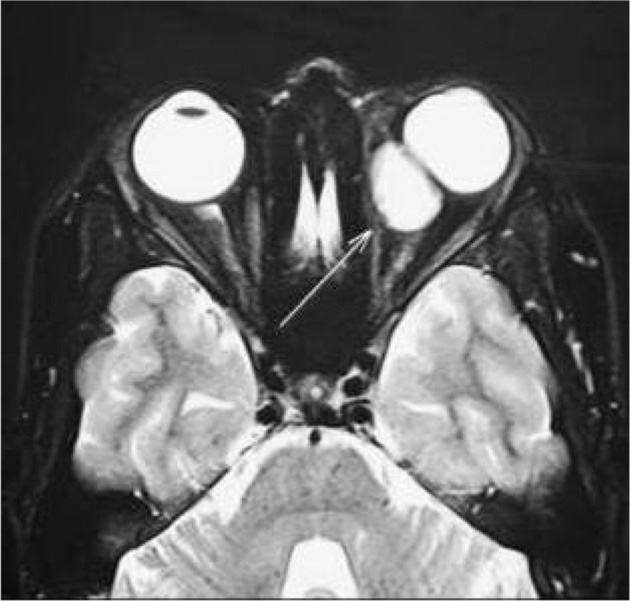
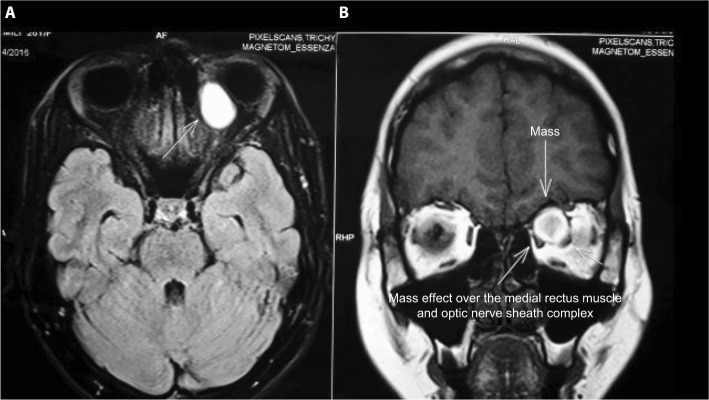
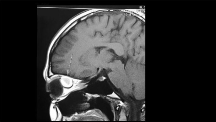
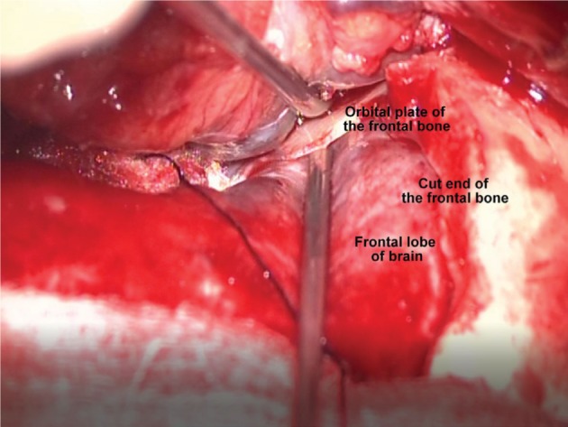
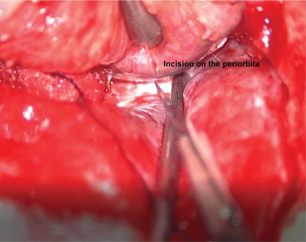
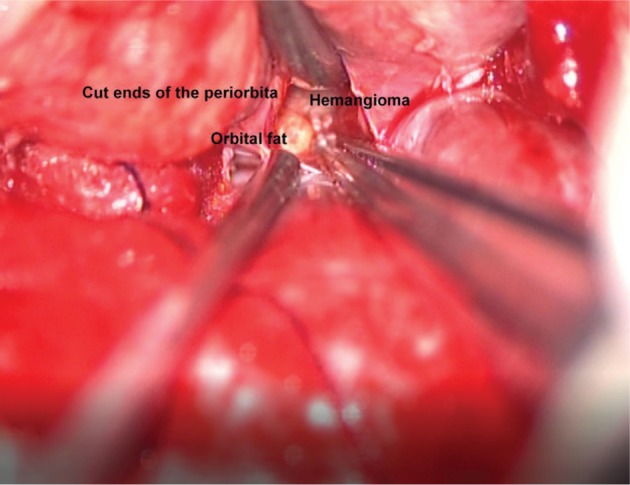
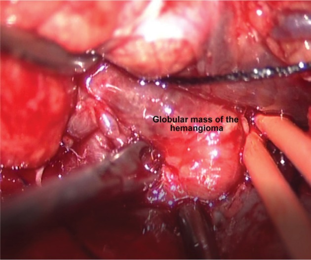
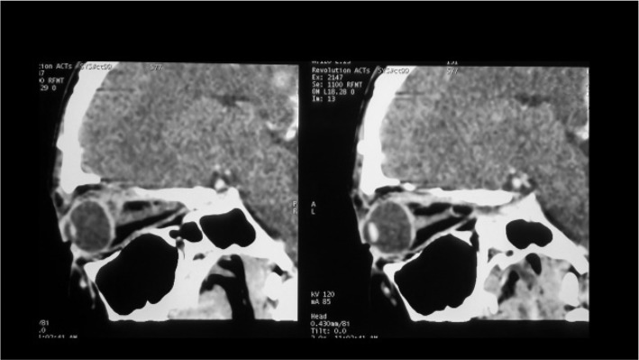
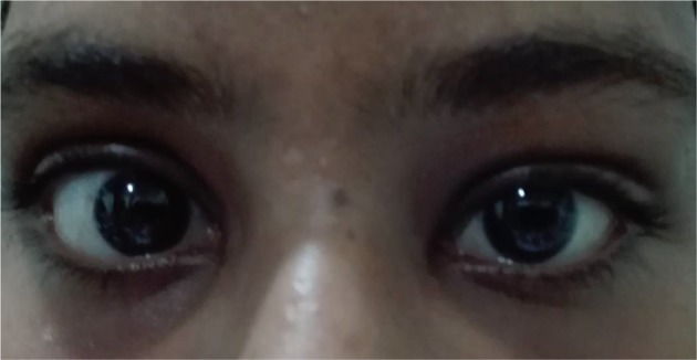
We report an unusual presentation of an orbital cavernous hemangioma in a 26-year-old female, who noted sudden redness and swelling of the left eye (LE) on waking up. At presentation, upper eyelid edema with periorbital ecchymosis and subconjunctival hemorrhage were noted in the LE. Although there was transient symptomatic relief with topical medications, blurring of vision developed in the LE. When seen 10 days later, the patient's LE showed axial proptosis. Magnetic resonance imaging revealed an intraconal soft tissue mass in the superomedial quadrant of the left orbit. Superior orbitotomy with mass excision was done; histopathological examination of the excised mass revealed a cavernous hemangioma. The patient had complete visual recovery following surgery. To our knowledge, an acute presentation of an orbital cavernous hemangioma with subconjunctival hemorrhage and periorbital ecchymosis has not previously been reported.
Keywords: cavernous hemangioma; ecchymosis; subconjunctival hemorrhage.
Conflict of interest statement
Disclosure The authors report no conflicts of interest in this work.
Figures
References
-
- Ansari SA, Mafee MF. Orbital cavernous hemangioma: role of imaging. Neuroimaging Clin N Am. 2005;15(1):137–158. - PubMed
-
- Arora V, Prat MC, Kazim M. Acute presentation of cavernous hemangioma of the orbit. Orbit. 2011;30(4):195–197. - PubMed
-
- Wills Eye Resident Case Series – Diagnosis and Discussion. [Accessed January 10, 2017]. Available from: https://www.reviewofophthalmology.com/.../wills-eye-resident-case-series....
Publication types
LinkOut - more resources
Full Text Sources
Other Literature Sources
