. 2017 Jul 18:8:1333.
doi: 10.3389/fmicb.2017.01333. eCollection 2017.

Fluorescence Imaging of Streptococcus pneumoniae with the Helix pomatia agglutinin (HPA) As a Potential, Rapid Diagnostic Tool

Affiliations

Fluorescence Imaging of Streptococcus pneumoniae with the Helix pomatia agglutinin (HPA) As a Potential, Rapid Diagnostic Tool

Mirian Domenech et al. Front Microbiol. .

Abstract

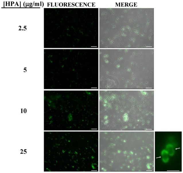
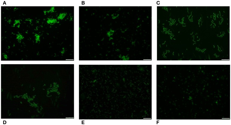
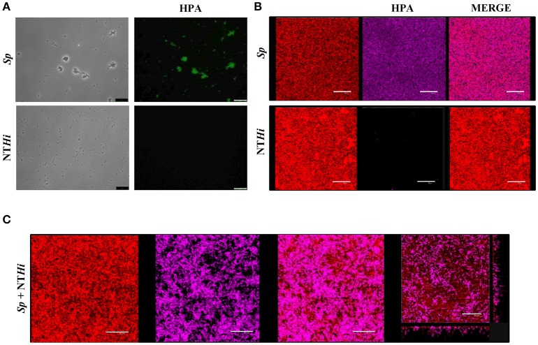
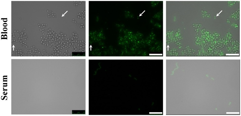
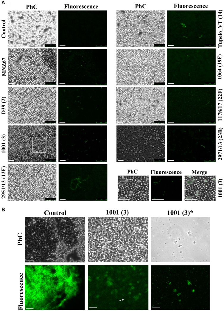
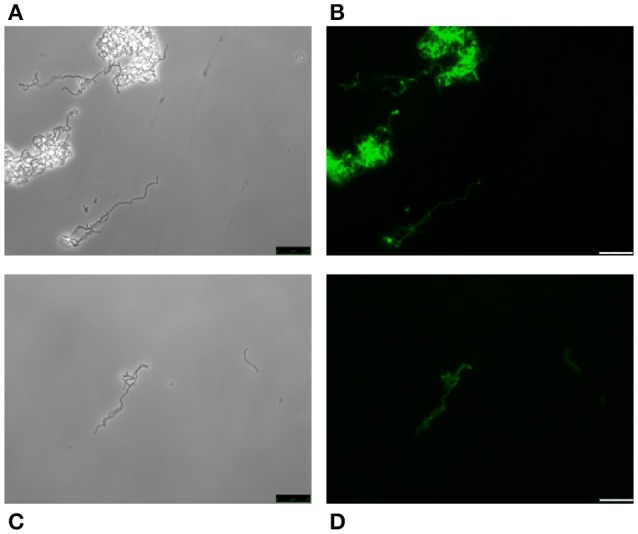
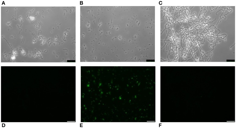
Streptococcus pneumoniae is a common human pathogen and a major causal agent of life-threatening infections that can either be respiratory or non-respiratory. It is well known that the Helix pomatia (edible snail) agglutinin (HPA) lectin shows specificity for terminal αGalNAc residues present, among other locations, in the Forssman pentasaccharide (αGalNAc1→3βGalNAc1→3αGal1→4βGal1→4βGlc). Based on experiments involving choline-independent mutants and different growth conditions, we propose here that HPA recognizes the αGalNAc terminal residues of the cell wall teichoic and lipoteichoic acids of S. pneumoniae. In addition, experimental evidence showing that pneumococci can be specifically labeled with HPA when growing as planktonic cultures as well as in mixed biofilms of S. pneumoniae and Haemophilus influenzae has been obtained. It should be underlined that pneumococci were HPA-labeled despite of the presence of a capsule. Although some non-pneumococcal species also bind the agglutinin, HPA-binding combined with fluorescence microscopy constitutes a suitable tool for identifying S. pneumoniae and, if used in conjunction with Gram staining and/or other suitable technique like antigen detection, it may potentially facilitate a fast and accurate diagnosis of pneumococcal infections.

Keywords: Forssman antigen; Streptococcus pneumoniae; binding lectins; fluorescence microscopy; teichoic acids.


Figures

Figure 1
Figure 1
Fluorescent labeling of the non-encapsulated S. pneumoniae strain R6 with HPA. Exponentially growing cultures of S. pneumoniae R6 in C+Y medium were incubated with the indicated concentrations of the lectin and observed for fluorescence (HCX PL FLUOTAR 40×/0.75 objective). Merges of fluorescence and phase-contrast images are also shown; bar = 25 μm. Enlarged view of two diplococci showing reduced fluorescence at the equatorial zone of growth (indicated by arrows; 63× objective). Bar = 2 μm.
Figure 2
Figure 2
HPA labeling of various streptococci in C+Y medium. First row: S. pneumoniae strains R6 (non-encapsulated) (A), D39 (serotype 2) (B), and P007 (serotype 3) (C). Second row: S. suis 298 (D), S. dysgalactiae subsp. equisimilis CECT 926 (E), and S. aureus type strain (F). Bar = 25 μm.
Figure 3
Figure 3
S. pneumoniae labeling with HPA in mixed biofilms. (A) Fluorescent labeling of the S. pneumoniae (Sp) strain R6 and the non-typeable H. influenzae (NTHi) 54997 with HPA. Biofilms formed by Sp R6 or NTHi 54997 (B), or both pathogens (C) were stained with a combination of SYTO 59 (red) and HPA conjugated to Alexa Fluor-488 (pink). An orthogonal projection of a CLSM image showing a representative region of the x–y plane over the depth of the biofilm in both x–z and y–z dimensions of the mixed biofilm is also shown at the bottom right part of the figure. Planktonic and biofilms cultures were incubated with HPA (25 μg/ml). Bar = 25 μm.
Figure 4
Figure 4
HPA labeling of S. pneumoniae in non-human blood and serum. The type 2 encapsulated S. pneumoniae strain D39 was diluted to 2.5 × 10⁶ cfu/ml into sheep blood or fetal bovine serum (63× objective). Cultures were incubated with HPA (25 μg/ml). Arrows point to pneumococcal cells. Bar = 25 μm.
Figure 5
Figure 5
HPA labeling of clinical pneumococcal isolates in human blood. The S. pneumoniae strains were diluted to 2.5 × 10⁶ cfu/ml into group O (A) and A (B) human blood (40× objective). Serotypes are indicated in parentheses. Strain MNZ67 is a non-encapsulated clinical isolate. In (A), the images shown at the bottom right are enlarged visions of the area marked with a rectangle in strain 1001. Control denotes non-infected blood samples. In (B), those labeled with an asterisk correspond to views of the supernatant of infected group A blood that had been centrifuged (1,000 × g; 1 min; room temperature) before HPA labeling. Samples were incubated with HPA (25 μg/ml). The arrow points to a pneumococcal diplococcus. Phc, phase-contrast micrograph; Bar = 25 μm.
Figure 6
Figure 6
HPA labeling of two choline-independent S. pneumoniae strains. Pneumococcal strains JY2190 (A,B) and P501 (C,D) were incubated in a chemically-defined medium (Cden) lacking any amino alcohol, and labeled with HPA (25 μg/ml). Bar = 25 μm.
Figure 7
Figure 7
Lack of HPA labeling in ethanolamine-containing streptococci. S. pneumoniae R6 cells were incubated for several generations in Cden medium containing ethanolamine (Cden-EA) (A,D). A portion of the culture then received choline chloride (5 μg/ml) and incubation proceeded at 37°C for 3 h (B,E). The S. mitis SK598 strain was grown in THY medium (C,F). The three cultures were incubated with HPA (25 μg/ml) (D–F). Bar = 25 μm.

Similar articles

Cited by

References

    1. Agger W. A., Maki D. G. (1978). Efficacy of direct Gram stain in differentiating staphylococci from streptococci in blood cultures positive for gram-positive cocci. J. Clin. Microbiol. 7, 111–113. - PMC - PubMed
    1. Ahmed S., Shapiro N. L., Bhattacharyya N. (2014). Incremental health care utilization and costs for acute otitis media in children. Laryngoscope 124, 301–305. 10.1002/lary.24190 - DOI - PubMed
    1. Amann R., Fuchs B. M. (2008). Single-cell identification in microbial communities by improved fluorescence in situ hybridization techniques. Nat. Rev. Micro. 6, 339–348. 10.1038/nrmicro1888 - DOI - PubMed
    1. Balsells E., Guillot L., Nair H., Kyaw M. H. (2017). Serotype distribution of Streptococcus pneumoniae causing invasive disease in children in the post-PCV era: a systematic review and meta-analysis. PLoS ONE 12:e0177113. 10.1371/journal.pone.0177113 - DOI - PMC - PubMed
    1. Bergström N., Jansson P. -E., Kilian M., Sørensen U. B. S. (2003). A unique variant of streptococcal group O-antigen (C-polysaccharide) that lacks phosphocholine. Eur. J. Biochem. 270, 2157–2162. 10.1046/j.1432-1033.2003.03569.x - DOI - PubMed