Review
. 2017 Jul 14:8:828.
doi: 10.3389/fimmu.2017.00828. eCollection 2017.

Reprogramming of Tumor-Associated Macrophages with Anticancer Therapies: Radiotherapy versus Chemo- and Immunotherapies

Affiliations
Review

Reprogramming of Tumor-Associated Macrophages with Anticancer Therapies: Radiotherapy versus Chemo- and Immunotherapies

Géraldine Genard et al. Front Immunol. .

Abstract

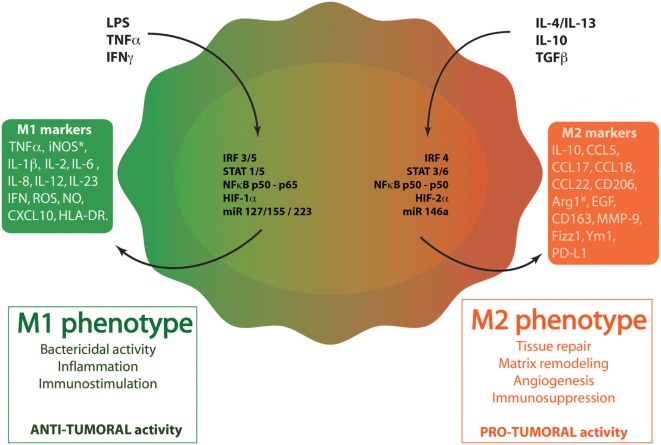
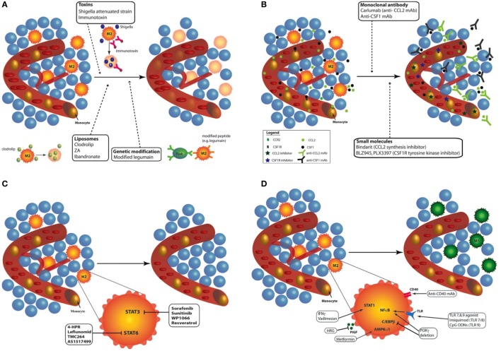
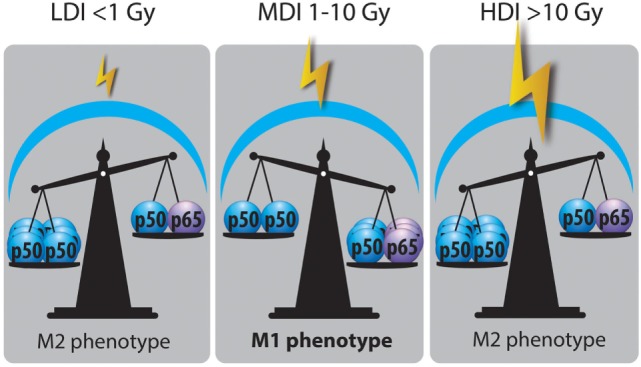
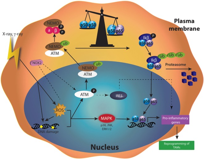
Tumor-associated macrophages (TAMs) play a central role in tumor progression, metastasis, and recurrence after treatment. Macrophage plasticity and diversity allow their classification along a M1-M2 polarization axis. Tumor-associated macrophages usually display a M2-like phenotype, associated with pro-tumoral features whereas M1 macrophages exert antitumor functions. Targeting the reprogramming of TAMs toward M1-like macrophages would thus be an efficient way to promote tumor regression. This can be achieved through therapies including chemotherapy, immunotherapy, and radiotherapy (RT). In this review, we first describe how chemo- and immunotherapies can target TAMs and, second, we detail how RT modifies macrophage phenotype and present the molecular pathways that may be involved. The identification of irradiation dose inducing macrophage reprogramming and of the underlying mechanisms could lead to the design of novel therapeutic strategies and improve synergy in combined treatments.

Keywords: chemotherapy; nuclear factor kappa B; polarization immunotherapy; radiotherapy; reactive oxygen species; reprogramming; tumor-associated macrophages.


Figures

Figure 1
Figure 1
Macrophage polarization. Through the binding to their respective receptors, M1 stimuli [lipopolysaccharide (LPS), tumor necrosis factor α (TNFα), and interferon γ (IFNγ)] trigger the activation of several transcription factors. These factors include interferon-regulatory factor/signal transducer and activator of transcription (IRF/STAT) family members (IRF3, IRF5, STAT1, and STAT5), the active nuclear factor kappa B (NFκB) heterodimer (p50–p65) and HIF1. miR127, miR 155, and miR223 also regulates M1 polarization. When polarized in M1-like phenotype, macrophages produce specific cytokines (TNFα, IL-1β, IL-2, IL-6, IL-12, IL-23, IFNγ), chemokines (CXCL10) and other molecules [reactive oxygen species (ROS), nitric oxide (NO), inducible nitric oxide synthase (iNOS), human leukocyte antigen-cell surface receptor (HLA-DR)]. M1 phenotype plays key roles in inflammation, immunostimulation and an antibacterial and antitumoral responses. M2 stimuli [IL-4, IL-13, IL-10, and transforming growth factor β (TGFβ)] bind to ILR4α, ILR10, or TGFβR to induce M2-like phenotype in macrophages. These stimuli activate several transcription factors: IRF/STAT family members (IRF4, STAT 3, and STAT6), the inhibitory NFκB homodimer (p50–p50) and HIF2. miR14a also influences M2 polarization. When polarized in M2-like phenotype, macrophages produce specific cytokines (IL-10), chemokines (CCL5, CCL17, CCL18, CCL22), and other proteins (CD163, CD206, Arg1, MMP-9, Fizz-1, Ym-1, and PD-L1). M2 macrophages exert diverse functions, such as tissue repair, matrix remodeling, angiogenesis, immunosuppression, and favor tumor growth.
Figure 2
Figure 2
Targeting tumor-associated macrophages (TAMs) with chemo- and immunotherapies. Different approaches have been proposed to modulate TAMs: (A) Depletion of TAMs: different kinds of treatments are available to destroy TAMs in tumor: toxins (Shigella flexneri attenuated strain or immunotoxin), liposome containing bisphosphonates [clodrolip, zoledronic acid (ZA) or ibandronate], and peptide modification to induce cytotoxic lymphocyte activation (e.g., legumain). Depletion of macrophages in tumor induced effective tumor regression in mouse and patients. (B) Inhibition of circulating monocyte recruitment into the tumor: two main recruitment effectors can be targeted to inhibit the recruitment of monocyte to the tumor site: CCL2/C–C chemokine receptor type 2 (CCR2) and colony-stimulating factor 1 (CSF-1)/CSF1-R. The use of monoclonal antibody against CCL2 (e.g., Carlumab) or CSF-1 inhibits tumor growth in mouse models and humans. Another way to prevent monocyte recruitment to the tumor site is the use of molecule targeting CCL2/CCR2 (e.g., bindarit) or CSF1/CSF1-R (e.g., BLZ945, PLX3397) pathways. (C) Blockade of M2 phenotype: the blockade of M2 phenotype can be achieved by targeting two main transcription factors: STAT3 (sorafenib, sunitinib, WP1066, and resveratrol) and STAT6 (4-HPR, leflunomid, TMX264, and AS1217499). All these inhibitors provide tumor regression and inhibited angiogenesis. (D) Enhanced activation of M1 macrophages or reprogramming of TAMs toward M1-like macrophages: TAM reprogramming into M1 macrophages can be achieved through the stimulation of STAT1 (IFNγ, vadimezan), AMPKα1 (metformin), or nuclear factor kappa B [toll-like receptor agonists such as imiquimod or CpG-ODNs; phosphoinositide 3-kinase (PI3Kγ) deletion]. The inhibition of placental growth factor (PlGF) (HRG) and C/EBPβ (PI3Kγ deletion) also leads to effective reprogramming of TAMs toward M1-like macrophages. Finally, by stimulating CD40, monoclonal antibodies (mAbs) against CD40 similarly reprogram TAMs from M2 phenotype to M1 macrophages.
Figure 3
Figure 3
Nuclear factor kappa B (NFκB) balance state after LDI, MDI, and HDI. Irradiation dose showed opposite effects on NFκB balance in macrophages: LDI or doses lower than 1 Gy did not modify the abundance of p50–p50 NFκB in macrophage nucleus after radiation. MDI or doses from 1 to 10 Gy induced a switch of NFκB balance from the inactive homodimer (p50–p50) to the active heterodimer (p50–p65), correlated with a reprogramming of tumor-associated macrophages (TAMs). HDI or doses higher than 10 Gy were not able to change the NFκB balance, skewing TAMs in a M2 phenotype (HDI, high doses of irradiation; MDI, moderated doses of irradiation; LDI, low doses of irradiation).
Figure 4
Figure 4
Molecular mechanisms activated after moderated doses of irradiation in tumor-associated macrophages (TAMs). Ionizing radiation (X-rays or γ-rays) induces DNA damage and elevated reactive oxygen species (ROS) content in cells. DNA repair machinery [such as ataxia telangiectasia mutated (ATM)] is activated by DNA damage and initiates the ubiquitination of NFκB essential modulator (NEMO), a subunit of the IKK complex. Therefore, ubiquinated NEMO can drive the activation of IKK complex in the cytoplasm. The degradation of IκB protein by the proteasome allows the release of p50–p65 nuclear factor kappa B (NFκB) in the cytoplasm. p50–p65 NFκB is then translocated into the nucleus and induces the transcription of pro-inflammatory genes, leading to the reprogramming of TAMs. ROS are also able to stimulate mitogen-activated protein kinase (MAPKs). Once phosphorylated, MAPKs also participate to the activation of NFκB and hence, to the transcription of pro-inflammatory genes.

References

    1. Mantovani A. Cancer: inflaming metastasis. Nature (2009) 457(7225):36–7.10.1038/457036b - DOI - PubMed
    1. Quail DF, Joyce JA. Microenvironmental regulation of tumor progression and metastasis. Nat Med (2013) 19(11):1423–37.10.1038/nm.3394 - DOI - PMC - PubMed
    1. Hanahan D, Weinberg RA. Hallmarks of cancer: the next generation. Cell (2011) 144(5):646–74.10.1016/j.cell.2011.02.013 - DOI - PubMed
    1. Schreiber RD, Old LJ, Smyth MJ. Cancer immunoediting: integrating immunity’s roles in cancer suppression and promotion. Science (2011) 331(6024):1565–70.10.1126/science.1203486 - DOI - PubMed
    1. Mittal D, Gubin MM, Schreiber RD, Smyth MJ. New insights into cancer immunoediting and its three component phases – elimination, equilibrium and escape. Curr Opin Immunol (2014) 27:16–25.10.1016/j.coi.2014.01.004 - DOI - PMC - PubMed