. 2017:2017:7617048.
doi: 10.1155/2017/7617048. Epub 2017 Jul 9.

NURR1 Downregulation Favors Osteoblastic Differentiation of MSCs

Affiliations

NURR1 Downregulation Favors Osteoblastic Differentiation of MSCs

Adriana Di Benedetto et al. Stem Cells Int. 2017.

Abstract

Mesenchymal stem cells (MSCs) have been identified in human dental tissues. Dental pulp stem cells (DPSCs) were classified within MSC family, are multipotent, can be isolated from adult teeth, and have been shown to differentiate, under particular conditions, into various cell types including osteoblasts. In this work, we investigated how the differentiation process of DPSCs toward osteoblasts is controlled. Recent literature data attributed to the nuclear receptor related 1 (NURR1), a still unclarified role in osteoblast differentiation, while NURR1 is primarily involved in dopaminergic neuron differentiation and activity. Thus, in order to verify if NURR1 had a role in DPSC osteoblastic differentiation, we silenced it during all the processes and compared the expression of the main osteoblastic markers with control cultures. Our results showed that the inhibition of NURR1 significantly increased the expression of osteoblast markers collagen I and alkaline phosphatase. Further, in long time cultures, the mineral matrix deposition was strongly enhanced in NURR1-silenced cultures. These results suggest that NURR1 plays a key role in switching DPSC differentiation toward osteoblasts rather than neuronal or even other cell lines. In conclusion, DPSCs represent a source of osteoblast-like cells and downregulation of NURR1 strongly prompted their differentiation toward the osteoblastogenesis process.


Figures

Figure 1
Figure 1
Expression of GFAP and nestin during osteogenic differentiation of DPSCs. Immunoblots show the protein expression trend of GFAP and nestin in DPSCs cultivated in osteogenic conditions for 4, 8, and 12 days. Both proteins were expressed during the first phases of osteogenic differentiation (4–8 days), but their expression dramatically dropped after 8 days of culture. Data are presented as means ± SE of 3 independent donors. P < 0.01 compared to T0. Statistics: unpaired Student's t-test.
Figure 2
Figure 2
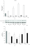
Effect of NURR1 short-interfering knockdown. (a) qPCR of DPSCs differentiated in osteogenic conditions and transfected with NURR1-specific siRNA (black bars) or scrambled sequences as control (white bars) showed the effective knockdown of NURR1 mRNA. A time course demonstrated that NURR1 expression remained downregulated along the culture. Expression was normalized to GAPDH. P < 0.01 compared to CTR. (b) NURR1 mRNA downregulation was confirmed and validated by Western blotting indicating that NURR1 protein was knocked down. Each graph represents means ± SE of 3 independent donors. P < 0.01 compared to CTR. Student's 𝑡-test was used for single comparison.
Figure 3
Figure 3
Effect of NURR1 downregulation on osteoblast markers. (a)-(b) qPCR performed on si-NURR1 or CTR cells showed that NURR1 downregulation significantly increased the expression of the two osteoblast markers ALP (8 days) (a) and Col1 (6–8 days) (b). Expression was normalized to GAPDH. P < 0.01 compared to CTR. (c) Immunoblotting confirmed that the expression of Col1 protein increased in NURR1 silenced cells relative to CTR cells (P < 0.01). Each graph represents means ± SE of 3 independent donors. Statistics: unpaired Student's t-test.
Figure 4
Figure 4
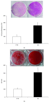
Effect of NURR1 downregulation on ALP and mineralization. (a) ALP histochemical assay (purple staining) performed on DPSCs transfected with NURR1-specific siRNA or scrambled sequences and maintained in osteogenic conditions for 7 days. The graph represents the quantification of positive staining as percentage compared to CTR (P < 0.01) and is representative for 3 independent donors. Data are presented as mean ± SEM. Student's 𝑡-test was used for single comparisons. (b) Mineral matrix deposition assayed by ARS (red staining) in siNURR1 and CTR cells after 21 days in osteogenic conditions. The graph shows the OD quantification of extracted dye from stained cell layers as percentage compared to CTR (P < 0.001) and is representative for 3 independent donors. Data are presented as mean ± SEM. Student's 𝑡-test was used for single comparisons.

References

    1. Gronthos S., Brahim J., Li W., et al. Stem cell properties of human dental pulp stem cells. Journal of Dental Research. 2002;81(8):531–535. doi: 10.1177/154405910208100806. - DOI - PubMed
    1. Gronthos S., Mankani M., Brahim J., Robey P. G., Shi S. Postnatal human dental pulp stem cells (DPSCs) in vitro and in vivo. Proceedings of the National Academy of Sciences of the United States of America. 2000;97(25):13625–13630. doi: 10.1073/pnas.240309797. - DOI - PMC - PubMed
    1. Sloan A. J., Smith A. J. Stem cells and the dental pulp: potential roles in dentine regeneration and repair. Oral Diseases. 2007;13(2):151–157. doi: 10.1111/j.1601-0825.2006.01346.x. - DOI - PubMed
    1. About I., Murray P. E., Franquin J. C., Remusat M., Smith A. J. The effect of cavity restoration variables on odontoblast cell numbers and dental repair. Journal of Dentistry. 2001;29(2):109–117. doi: 10.1016/S0300-5712(00)00067-1. - DOI - PubMed
    1. Murray P. E., About I., Franquin J. C., Remusat M., Smith A. J. Restorative pulpal and repair responses. Journal of the American Dental Association (1939) 2001;132(4):482–491. - PubMed

LinkOut - more resources