

Brain-Derived Neurotrophic Factor Attenuates Septic Myocardial Dysfunction via eNOS/NO Pathway in Rats
- PMID: 28770018
- PMCID: PMC5523440
- DOI: 10.1155/2017/1721434
Brain-Derived Neurotrophic Factor Attenuates Septic Myocardial Dysfunction via eNOS/NO Pathway in Rats
Abstract
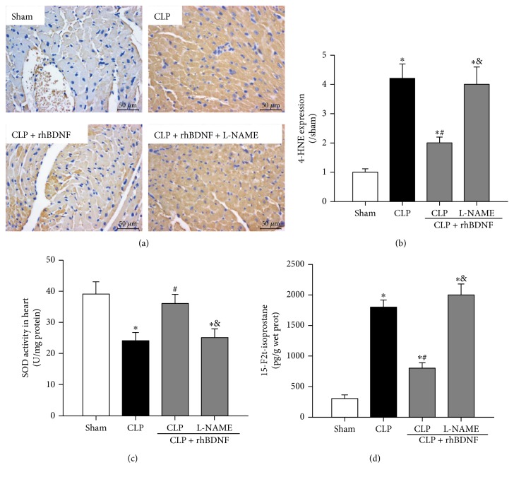
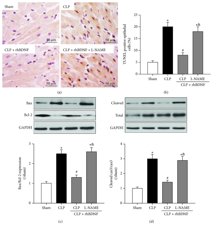
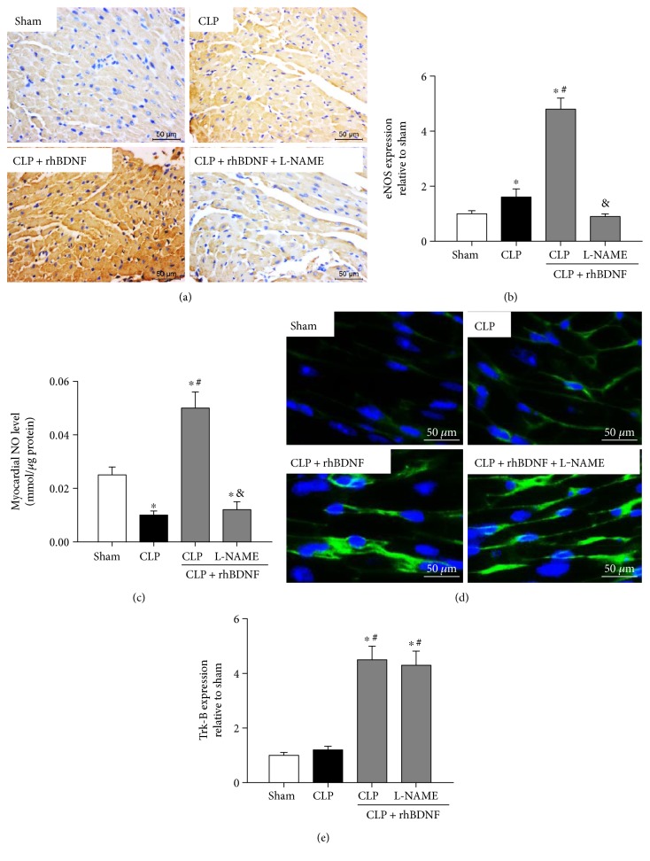
Sepsis-induced myocardial dysfunction increases mortality in sepsis, yet the underlying mechanism is unclear. Brain-derived neurotrophic factor (BDNF) has been found to enhance cardiomyocyte function, but whether BDNF has a beneficial effect against septic myocardial dysfunction is unknown. Septic shock was induced by cecal ligation and puncture (CLP). BDNF was expressed in primary cardiomyocytes, and its expression was significantly reduced after sepsis. In rats with sepsis, a sharp decline in survival was observed after CLP, with significantly reduced cardiac BDNF expression, enhanced myocardial fibrosis, elevated oxidative stress, increased myocardial apoptosis, and decreased endothelial nitric oxide (NO) synthase (eNOS) and NO. Supplementation with recombined BDNF protein (rhBDNF) enhanced myocardial BDNF and increased survival rate with improved cardiac function, reduced oxidative stress, and myocardial apoptosis, which were associated with increased eNOS expression, NO production, and Trk-B, a BDNF receptor. Pretreatment with NOS inhibitor, N (omega)-nitro-L-arginine methyl ester, abolished the abovementioned BDNF cardioprotective effects without affecting BDNF and Trk-B. It is concluded that BDNF protects the heart against septic cardiac dysfunction by reducing oxidative stress and apoptosis via Trk-B, and it does so through activation of eNOS/NO pathway. These findings provide a new treatment strategy for sepsis-induced myocardial dysfunction.
Figures


Similar articles
-
N-acetylcysteine attenuates myocardial dysfunction and postischemic injury by restoring caveolin-3/eNOS signaling in diabetic rats.Cardiovasc Diabetol. 2016 Oct 12;15(1):146. doi: 10.1186/s12933-016-0460-z. Cardiovasc Diabetol. 2016. PMID: 27733157 Free PMC article.
-
Myocardial caspase-3 and NF-κB activation promotes calpain-induced septic apoptosis: The role of Akt/eNOS/NO pathway.Life Sci. 2019 Apr 1;222:195-202. doi: 10.1016/j.lfs.2019.02.048. Epub 2019 Feb 23. Life Sci. 2019. PMID: 30807754
-
Brain-derived neurotrophic factor attenuates doxorubicin-induced cardiac dysfunction through activating Akt signalling in rats.J Cell Mol Med. 2017 Apr;21(4):685-696. doi: 10.1111/jcmm.13012. Epub 2016 Nov 7. J Cell Mol Med. 2017. PMID: 28098423 Free PMC article.
-
[Impact of nitric oxide synthase 3 on myocardial dysfunction in sepsis].Masui. 2008 Mar;57(3):294-301. Masui. 2008. PMID: 18340997 Review. Japanese.
-
Molecular events in the cardiomyopathy of sepsis.Mol Med. 2008 May-Jun;14(5-6):327-36. doi: 10.2119/2007-00130.Flierl. Mol Med. 2008. PMID: 18256728 Free PMC article. Review.
Cited by
-
An Integrative Review of Brain-Derived Neurotrophic Factor and Serious Cardiovascular Conditions.Nurs Res. 2020 Sep/Oct;69(5):376-390. doi: 10.1097/NNR.0000000000000454. Nurs Res. 2020. PMID: 32555009 Free PMC article. Review.
-
BDNF-induced BDNF release: A virtuous loop for the cardioprotective effects of exercise in post-ischemic heart failure.Int J Cardiol Heart Vasc. 2025 Jan 24;56:101623. doi: 10.1016/j.ijcha.2025.101623. eCollection 2025 Feb. Int J Cardiol Heart Vasc. 2025. PMID: 39927379 Free PMC article. No abstract available.
-
Aerosol inhalation of a hydrogen-rich solution restored septic renal function.Aging (Albany NY). 2019 Dec 16;11(24):12097-12113. doi: 10.18632/aging.102542. Epub 2019 Dec 16. Aging (Albany NY). 2019. PMID: 31841441 Free PMC article.
-
Protective effects of Dioscin against sepsis-induced cardiomyopathy via regulation of toll-like receptor 4/MyD88/p65 signal pathway.Immun Inflamm Dis. 2024 May;12(5):e1229. doi: 10.1002/iid3.1229. Immun Inflamm Dis. 2024. PMID: 38775678 Free PMC article.
-
Obese mice exposed to psychosocial stress display cardiac and hippocampal dysfunction associated with local brain-derived neurotrophic factor depletion.EBioMedicine. 2019 Sep;47:384-401. doi: 10.1016/j.ebiom.2019.08.042. Epub 2019 Sep 3. EBioMedicine. 2019. PMID: 31492565 Free PMC article.
References
-
- Bone R. C., Balk R. A., Cerra F. B., et al. Definitions for sepsis and organ failure and guidelines for the use of innovative therapies in sepsis. The ACCP/SCCM Consensus Conference Committee. American College of Chest Physicians/Society of Critical Care Medicine. Chest. 1992;101(6):1644–1655. doi: 10.1378/chest.101.6.1644. - DOI - PubMed
MeSH terms
Substances
LinkOut - more resources
Full Text Sources
Other Literature Sources
Medical
Miscellaneous
