Review
. 2017:2017:7027380.
doi: 10.1155/2017/7027380. Epub 2017 Jul 9.

Potential Roles of Exosomal MicroRNAs as Diagnostic Biomarkers and Therapeutic Application in Alzheimer's Disease

Affiliations
Review

Potential Roles of Exosomal MicroRNAs as Diagnostic Biomarkers and Therapeutic Application in Alzheimer's Disease

Jian-Jiao Chen et al. Neural Plast. 2017.

Abstract

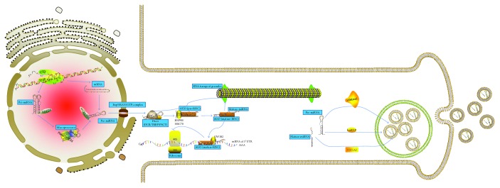
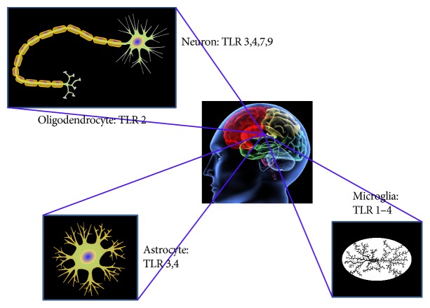
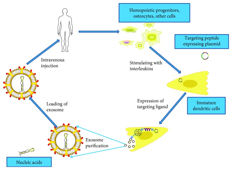
Exosomes are bilipid layer-enclosed vesicles derived from endosomes and are released from neural cells. They contain a diversity of proteins, mRNAs, and microRNAs (miRNAs) that are delivered to neighboring cells and/or are transported to distant sites. miRNAs released from exosomes appear to be associated with multiple neurodegenerative conditions linking to Alzheimer's disease (AD) which is marked by hyperphosphorylated tau proteins and accumulation of Aβ plaques. Exciting findings reveal that miRNAs released from exosomes modulate the expression and function of amyloid precursor proteins (APP) and tau proteins. These open up the possibility that dysfunctional exosomal miRNAs may influence AD progression. In addition, it has been confirmed that the interaction between miRNAs released by exosomes and Toll-like receptors (TLR) initiates inflammation. In exosome support-deprived neurons, exosomal miRNAs may regulate neuroplasticity to relieve neurological damage. In this review, we summarize the literature on the function of exosomal miRNAs in AD pathology, the potential of these miRNAs as diagnostic biomarkers in AD, and the use of exosomes in the delivery of miRNAs which may lead to major advances in the field of macromolecular drug delivery.


Figures

Figure 1
Figure 1
The biogenesis pathway of exosomal miRNA and composition of exosome. Canonical miRNAs are initially transcribed from long (41 kb) endogenous precursors called primary miRNAs that are driven by RNA pol II promoters and cleaved by microprocessor, a multiprotein complex formed by the RNase type III Drosha (the Drosophila homolog of RNASEN in humans) and the protein DGCR8 (Di George Syndrome Critical Region 8). A hairpin structure (now known as pre-miRNA) with 60–80 nucleotides in length, which bears a two-nucleotide overhang at the 3′ end that is a mark left by the Drosha processing, is released in this step. Within neuronal nuclei, pri- and pre-miRNA may be stabilized by 3′-terminal adenylation performed by PAPD4. After recognizing the precursors by their overhangs, GTPase-dependent Ran-Exp5 complex exports the pre-miRNAs out of the nucleus to the cytoplasm. An alternative pathway needs splicing out of the miRNAs from introns located in other genes, then further lariat processing, and finally proper folding into a pre-miRNA structure. In the cytoplasm, cleavage of the pre-miRNAs takes place once they have been loaded onto the Dicer-TRBP (TAR-RNA binding protein) complex, which removes the loop from the pre-miRNA to produce a dsRNA duplex that contains both the mature miRNA (or leader strand) and the so-called passenger strand. The Dicer-TRBP complex rapidly transfers the duplex to the miRISC (miRNA-RNAi-induced silencing complex), which contains AGO (Argonaute proteins) as its core. The HSP90/HSC70 chaperone complex participates in the process. In the miRISC, the passenger strand is degraded by an unidentified mechanism, leaving a mature miRISC loaded with a fully mature single-stranded miRNA (19–22 nt) to bind canonically to non-fully complementary mRNA targets at their 3′ untranslated regions (UTR). Alternatively, pri-miRNAs and miRNA may be loaded with proteins of RNA transport granules. These molecules are then transported to specific neuronal compartments, where mature or precursor miRNAs are enveloped in exosomes to be released elsewhere. Mature miRNAs are sorted into exosomes via four potential mechanisms: (1) the neutral sphingomyelinase 2- (nSMase2-) dependent pathway; (2) the miRNA motif and sumoylated heterogeneous nuclear ribonucleoprotein- (hnRNP-) dependent pathway; (3) the 3′-end of the miRNA sequence-dependent pathway; and (4) the miRNA-induced silencing complex- (miRISC-) related pathway (not shown).
Figure 2
Figure 2
Neural cell types and their Toll-like receptor (TLR) expression. Protein profile for TLR3, 4, 7, and 9 has been reported in different neural phenotypes from humans; only TLR2 protein profile is detected in human oligodendrocytes; TLR3-4 protein accumulation is found in human astrocytes; human microglia contains TLR1–4 protein profile.
Figure 3
Figure 3
Schematic representation of production, harvest, and readministration of engineering modified exosomes for gene delivery. To acquire enough immunologically inert exosomes, harvest cells like hemopoietic progenitors and osteocytes are used as the source cell. As immature dendritic cells produce a lot of exosomes devoid of T-cell activators such as MHC-II and CD86, it could be selected as the source cell. Targeting peptides expressing plasmids (e.g., RVG (rabies virus glycoprotein)) were transfected into the source cells to get exosomes with the ability of specifically binding to neural cells. Therapeutic nucleic acids can be introduced into the modified exosomes by electroporation method. Following intravenous injection, exosomes encapsulating nucleic acids could be found within the central nervous system.

References

    1. Bennett D. A., Yu L., Yang J., Srivastava G. P., Aubin C., De Jager P. L. Epigenomics of Alzheimer’s disease. Translational Research. 2015;165(1):200–220. doi: 10.1016/j.trsl.2014.05.006. - DOI - PMC - PubMed
    1. Villemagne V. L., Burnham S., Bourgeat P., et al. Amyloid beta deposition, neurodegeneration, and cognitive decline in sporadic Alzheimer’s disease: a prospective cohort study. Lancet Neurology. 2013;12(4):357–367. doi: 10.1016/S1474-4422(13)70044-9. - DOI - PubMed
    1. Liu C. G., Song J., Zhang Y. Q., Wang P. C. MicroRNA-193b is a regulator of amyloid precursor protein in the blood and cerebrospinal fluid derived exosomal microRNA-193b is a biomarker of Alzheimer’s disease. Molecular Medicine Reports. 2014;10(5):2395–2400. doi: 10.3892/mmr.2014.2484. - DOI - PubMed
    1. Lugli G., Cohen A. M., Bennett D. A., et al. Plasma exosomal miRNAs in persons with and without Alzheimer disease: altered expression and prospects for biomarkers. PloS One. 2015;10(10, article e0139233) - PMC - PubMed
    1. Gui Y., Liu H., Zhang L., Lv W., Hu X. Altered microRNA profiles in cerebrospinal fluid exosome in Parkinson disease and Alzheimer disease. Oncotarget. 2015;6(35):37043–37053. doi: 10.18632/oncotarget.6158. - DOI - PMC - PubMed