. 2017 Jul 13;3(1):92-109.
doi: 10.1016/j.chempr.2017.05.016.

Nanoparticle-Based Receptors Mimic Protein-Ligand Recognition

Affiliations

Nanoparticle-Based Receptors Mimic Protein-Ligand Recognition

Laura Riccardi et al. Chem. .

Abstract

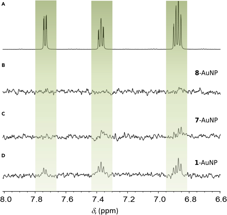
The self-assembly of a monolayer of ligands on the surface of noble-metal nanoparticles dictates the fundamental nanoparticle's behavior and its functionality. In this combined computational-experimental study, we analyze the structure, organization, and dynamics of functionalized coating thiols in monolayer-protected gold nanoparticles (AuNPs). We explain how functionalized coating thiols self-organize through a delicate and somehow counterintuitive balance of interactions within the monolayer itself and with the solvent. We further describe how the nature and plasticity of these interactions modulate nanoparticle-based chemosensing. Importantly, we found that self-organization of coating thiols can induce the formation of binding pockets in AuNPs. These transient cavities can accommodate small molecules, mimicking protein-ligand recognition, which could explain the selectivity and sensitivity observed for different organic analytes in NMR chemosensing experiments. Thus, our findings advocate for the rational design of tailored coating groups to form specific recognition binding sites on monolayer-protected AuNPs.

Keywords: AuNP; NMR chemosensing; NMR relaxation; NOE; SDG3: Good health and well-being; molecular dynamics of gold nanoparticles; molecular recognition; molecular simulations; monolayer-protected nanoparticles; self-organization.


Figures

None
Graphical abstract
Scheme 1
Scheme 1
The NMR Chemosensing Machinery for Analyte Detection Upper: the monolayer-protected nanoparticles (ligands 1 and 2) and analytes (36) investigated in early studies. Lower: nanoparticle-coating thiols (1 and 710) used in this study and the solubility of representative samples in CHCl3 and water.
Figure 1
Figure 1
NMR Spectra of 1-, 7-, and 8-AuNP in D2O and CDCl3 Signals from the OEG portion of the coating thiols are highlighted in blue, and signals from the alkyl portions are highlighted in red. Symbols are as follows: *, residual water signal; and °, impurities.
Figure 2
Figure 2
NMR Spectra (A) 1H NMR subspectrum of 5 mM sodium salicylate (3) in D2O. (B) NOE-pumping subspectrum of the same sample in the presence of 8-AuNP. (C) NOE-pumping subspectrum of the same sample in the presence of 7-AuNP. (D) NOE-pumping subspectrum of the same sample in the presence of 1-AuNP. Conditions: [AuNP] = 15 μM, carbonate buffer 20 mM, pD = 10, 298 K. See also Supplemental Information Section S5 and Figures S33–S35.
Figure 3
Figure 3
Comparison of Experimental and Calculated T1 Values of 13C Nuclei along the Chains of 1-AuNPs Left: 1-AuNPs dissolved in water. Right: 1-AuNPs dissolved in chloroform. T = 298 K. 13C Larmor frequency = 125.75 MHz. See also Supplemental Information Section S11.6 and Figure S74.
Figure 4
Figure 4
Shape and Solvation of AuNPs (A and B) Probability distributions of (A) radius of gyration (Rg) and (B) moments of inertia (I, shown as box-and-whisker plots) of 1-, 7-, 8-, 9-, and 10-AuNPs in water and chloroform. The average nanoparticle eccentricity e = (1 − Imin/Iavg), where e = 0 for a sphere and 1 for a prolate spheroid, is also reported (SD = 0.2 except for * and #, where it is 0.3 and 0.4, respectively). (C) Distribution of the solvent molecules and the gold and sulfur atoms (in the inset in yellow and orange, respectively) from the center of mass of the Au144 core. (D) Representative snapshots of the AuNPs and solvent molecules within 1 nm of the gold atoms (wires connect carbon atoms C4–C7 closer than 0.8 nm). See also Supplemental Information Section S11.2 and Figure S70.
Figure 5
Figure 5
HB Interactions Average number of ligand-ligand and ligand-water HBs during MD simulations and the decomposition of this number for the different coating thiol atoms (ordered top to bottom from the outer to the inner part of the coating ligand). For NH/O, NH is present in 1-AuNP and O is present in 7- and 8-AuNPs.
Figure 6
Figure 6
Structure of the Monolayer (A) Amide group atoms closer than 0.4 nm are connected by wires, and solvent molecules within 1 nm of the Au144 core are shown. The Au144 core is shown as a gold surface, and sulfur atoms are shown as orange spheres. (B) Example of the HB network for 1-AuNP in water. (C) Identification of pockets on one snapshot of 1-AuNP in water. (D) Superposition of the docking pose of salicylate in 1-AuNP and in the LysR-type transcription factor (PDB: 2Y7K). (E) Characterization of pockets for 1-AuNP in water. Magenta spheres indicate the “deep cavity” pockets (left), and yellow spheres indicate the “OEG sinking” pockets (right). Gray, green, and cyan surfaces identify the alkyl, amide, and OEG regions, respectively. (F) Time evolution and lifetime of a stable pocket formed in 1-AuNP in water. Blue color for the open pocket and black for the closed pocket. See also Supplemental Information Sections S11.3–S11.4 and S11.7–S11.8 and Figures S71–72, S75, and S76.
Figure 7
Figure 7
Interactions between Analyte and AuNP (A) 1H reference spectrum of 20 mM sodium salicylate and 10 mM 1-AuNP in D2O carbonate buffer (pD = 10). (B) 1D NOESY spectrum of the same sample obtained with selective excitation of the salicylate resonances (6–9 ppm) and 300-ms mixing time. Signals of the nanoparticle highlight a negative NOE regime (slow tumbling). (C) Number of total 1H-1H contacts, color coded in intervals of 1 Å, between docked salicylate and 1H-bearing atoms of 1-AuNP (exchangeable NH omitted). The bars have been sorted according to the chemical shifts of the parent atoms. See also Supplemental Information Section S12 and Figure S78.
Figure 8
Figure 8
1-AuNP-Analyte Interactions (A) NOE-pumping spectra resulting from a solution of 15 μM 1-AuNP with either 5 mM salicylate (left) or 5 mM 4-hydroxybenzoate (right). Mixing time is 1.2 s. The emergence of a signal only for salicylate is well rationalized by the MD results outlined in (B) (see text for details). (B) During MD simulations, 1-AuNP-analyte binding events were defined when the minimum intermolecular proton-proton distance was less than 0.4 nm. The binding events were sorted by their binding residence time (x coordinate) and plotted against the minimum distance between the analyte and the Au144 core (depth of penetration in the monolayer, y coordinate). The rotational correlation time (τc) of 1-AuNP is also reported as a visual guideline. (C) Distribution of the analytes in the monolayer taken from MD snapshots every 25 ns. Purple analytes lie at max 1 nm from the gold core, and green analytes lie between 1 and 1.5 nm. (D) Binding event of salicylate to 1-AuNP. See also Supplemental Information Section S11.9, Figure S77, and Movie S1.

References

    1. Gentilini C., Franchi P., Mileo E., Polizzi S., Lucarini M., Pasquato L. Formation of patches on 3D SAMs driven by thiols with immiscible chains observed by ESR spectroscopy. Angew. Chem. Int. Ed. 2009;48:3060–3064. - PubMed
    1. Stewart A., Zheng S., McCourt M.R., Bell S.E.J. Controlling assembly of mixed thiol monolayers on silver nanoparticles to tune their surface properties. ACS Nano. 2012;6:3718–3726. - PMC - PubMed
    1. Shaw C.P., Fernig D.G., Lévy R. Gold nanoparticles as advanced building blocks for nanoscale self-assembled systems. J. Mater. Chem. 2011;21:12181–12187.
    1. Della Sala F., Kay E.R. Reversible control of nanoparticle functionalization and physicochemical properties by dynamic covalent exchange. Angew. Chem. Int. Ed. 2015;54:4187–4191. - PMC - PubMed
    1. Badia A., Singh S., Demers L., Cuccia L., Brown G.R., Lennox R.B. Self-assembled monolayers on gold nanoparticles. Chem. A. Eur. J. 1996;2:359–363.

LinkOut - more resources