. 2017 Sep;264(9):1956-1967.
doi: 10.1007/s00415-017-8581-1. Epub 2017 Aug 2.

Severity of traumatic brain injury correlates with long-term cardiovascular autonomic dysfunction

Affiliations

Severity of traumatic brain injury correlates with long-term cardiovascular autonomic dysfunction

Max J Hilz et al. J Neurol. 2017 Sep.

Abstract

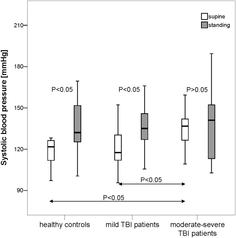
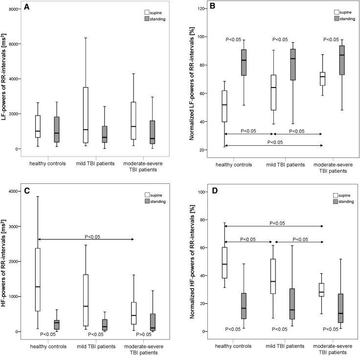
After traumatic brain injury (TBI), central autonomic dysfunction might contribute to long-term increased mortality rates. Central autonomic dysfunction might depend on initial trauma severity. This study was performed to evaluate differences in autonomic modulation at rest and upon standing between patients with a history of mild TBI (post-mild-TBI patients), moderate or severe TBI (post-moderate-severe-TBI patients), and healthy controls. In 20 post-mild-TBI patients (6-78 months after TBI), age-matched 20 post-moderate-severe-TBI patients (6-94 months after TBI) and 20 controls, we monitored respiration, RR intervals (RRI) and systolic blood pressure (BPsys) at supine rest and upon standing. We determined mainly sympathetic low (LF) and parasympathetic high (HF) frequency powers of RRI fluctuations, sympathetically mediated LF-BPsys powers, LF/HF-RRI ratios, normalized (nu) LF-RRI and HF-RRI powers, and compared data between groups, at rest and upon standing (ANOVA with post hoc testing). We correlated autonomic parameters with initial Glasgow Coma Scale (GCS) scores (Spearman test; significance: p < 0.05). Supine BPsys and LFnu-RRI powers were higher while HFnu-RRI powers were lower in post-moderate-severe-TBI patients than post-mild-TBI patients and controls. LFnu-RRI powers were higher and HFnu-RRI powers were lower in post-mild-TBI patients than controls. Upon standing, only post-mild-TBI patients and controls increased LF-BPsys powers and BPsys and decreased HF-RRI powers. GCS scores correlated positively with LFnu-RRI powers, LF/HF-RRI ratios, and inversely with HFnu-RRI powers, at standing position. More than 6 months after TBI, there is autonomic dysfunction at rest and upon standing which is more pronounced after moderate-severe than mild TBI and in part correlates with initial trauma severity.

Keywords: Autonomic dysfunction; Differences in severity; Orthostatic challenge; Traumatic brain injury.


Conflict of interest statement

Conflicts of interest

On behalf of all authors, the corresponding author states that there is no conflict of interest.

Ethical standards

The Institutional Review Board (IRB) at the New York University and the ethics committee of the University of Erlangen-Nuremberg, Germany, had approved the study and written informed consent was obtained from all participants according to the declaration of Helsinki (2000) of the World Medical Association.

Figures

Fig. 1
Fig. 1
Systolic blood pressure (BPsys) in supine and standing positions in 20 patients with a history of mild TBI, 20 patients with a history of moderate or severe TBI (moderate–severe TBI) and 20 healthy controls. In supine position, BPsys was significantly higher in moderate–severe TBI patients than in mild-TBI patients or controls (P < 0.05). Upon standing, healthy controls and mild-TBI patients significantly increased BPsys while moderate–severe TBI patients did not change their BPsys. Data are presented as box plots. The top of the box represents the 75th percentile (upper quartile), the bottom of the box represents the 25th percentile (lower quartile), the line in the middle represents the 50th percentile (median). The ends of the whiskers represent the highest and lowest values that are not outliers or extreme values. White boxes illustrate supine values, and gray boxes illustrate standing values
Fig. 2
Fig. 2
Absolute and normalized low-frequency powers of RR intervals (LF-RRI) and high-frequency powers of RR intervals (HF-RRI) in supine and standing positions in 20 patients with a history of mild TBI, 20 patients with a history of moderate or severe TBI (moderate–severe TBI) and 20 healthy controls. At supine rest, normalized RRI-LF powers (LFnu-RRI powers) were significantly higher while normalized RRI-HF powers (HFnu-RRI powers) were significantly lower in the moderate–severe TBI patients group than in the patients with a history of mild TBI or the healthy controls (P < 0.05). In mild-TBI patients, LFnu-RRI powers again were significantly higher and HFnu-RRI powers were lower than in controls. In moderate–severe TBI patients, the parasympathetically mediated HF-RRI powers were significantly lower than in healthy controls. Upon standing up, all groups significantly decreased normalized HF-RRI powers, and significantly increased normalized LF-RRI powers, LF-RRI powers did not change in any group (P > 0.05). Healthy controls and mild-TBI patients significantly decreased HF-RRI powers, while moderate–severe TBI patients did not change their HF-RRI powers upon standing-up. a Absolute LF powers of RR intervals; b normalized LF powers of RR intervals; c absolute HF powers of RR intervals; d normalized HF powers of RR intervals. Data are presented as box plots. The top of the box represents the 75th percentile (upper quartile), the bottom of the box represents the 25th percentile (lower quartile), the line in the middle represents the 50th percentile (median). The ends of the whiskers represent the highest and lowest values that are not outliers or extreme values. White boxes illustrate supine values, and gray boxes illustrate standing values

References

    1. Abaji JP, Curnier D, Moore RD, Ellemberg D. Persisting effects of concussion on heart rate variability during physical exertion. J Neurotrauma. 2015;33:811–817. doi: 10.1089/neu.2015.3989. - DOI - PubMed
    1. Annegers JF, Hauser WA, Coan SP, Rocca WA. A population-based study of seizures after traumatic brain injuries. N Engl J Med. 1998;338:20–24. doi: 10.1056/NEJM199801013380104. - DOI - PubMed
    1. Benarroch EE. The central autonomic network: functional organization, dysfunction, and perspective. Mayo Clin Proc. 1993;68:988–1001. doi: 10.1016/S0025-6196(12)62272-1. - DOI - PubMed
    1. Black M, Graham DI. Sudden unexplained death in adults caused by intracranial pathology. J Clin Pathol. 2002;55:44–50. doi: 10.1136/jcp.55.1.44. - DOI - PMC - PubMed
    1. Bondanelli M, De Marinis L, Ambrosio MR, Monesi M, Valle D, Zatelli MC, Fusco A, Bianchi A, Farneti M, Degli EC. Occurrence of pituitary dysfunction following traumatic brain injury. J Neurotrauma. 2004;21:685–696. doi: 10.1089/0897715041269713. - DOI - PubMed

MeSH terms