. 2017 Aug 3:8:14130.
doi: 10.1038/ncomms14130.

Identification and characterization of a novel botulinum neurotoxin

Affiliations

Identification and characterization of a novel botulinum neurotoxin

Sicai Zhang et al. Nat Commun. .

Abstract

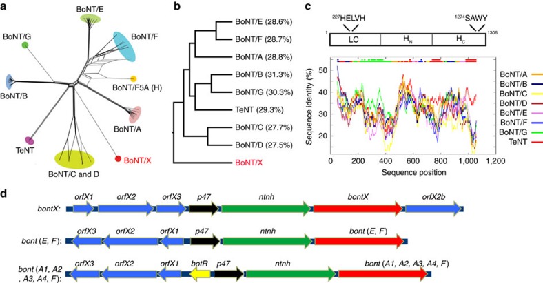
Botulinum neurotoxins are known to have seven serotypes (BoNT/A-G). Here we report a new BoNT serotype, tentatively named BoNT/X, which has the lowest sequence identity with other BoNTs and is not recognized by antisera against known BoNTs. Similar to BoNT/B/D/F/G, BoNT/X cleaves vesicle-associated membrane proteins (VAMP) 1, 2 and 3, but at a novel site (Arg66-Ala67 in VAMP2). Remarkably, BoNT/X is the only toxin that also cleaves non-canonical substrates VAMP4, VAMP5 and Ykt6. To validate its activity, a small amount of full-length BoNT/X was assembled by linking two non-toxic fragments using a transpeptidase (sortase). Assembled BoNT/X cleaves VAMP2 and VAMP4 in cultured neurons and causes flaccid paralysis in mice. Thus, BoNT/X is a novel BoNT with a unique substrate profile. Its discovery posts a challenge to develop effective countermeasures, provides a novel tool for studying intracellular membrane trafficking, and presents a new potential therapeutic toxin for modulating secretions in cells.


Conflict of interest statement

Boston Children’s Hospital has filed a provisional patent application (U.S. 62/360,239) for medical use of BoNT/X, with P.S., S.Z. and M.D. as inventors.

Figures

Figure 1
Figure 1. Identification of BoNT/X.
(a) A phylogenetic split network covering all BoNT serotypes, subtypes, mosaic toxins and related tetanus neurotoxin (TeNT) illustrates their potential evolutionary relationships, as well as conflicts arising from e.g. chimerisms, based on their protein sequences. BoNT/X is highlighted in red. An enlarged version of this panel is shown in Supplementary Fig. 1, with the sequence access number for each toxin gene noted. (b) A phylogenic tree of the protein sequence alignment for BoNT/A-G, TeNT and BoNT/X, analysed by the ClustalW method. The percentages of sequence identity between each toxin and BoNT/X are noted. (c) Upper panel: a schematic drawing of the three domains of BoNT/X, with conserved protease motif in the LC and the ganglioside binding motif in the HC noted. Lower panel: analysis using a sliding sequence comparison window demonstrated that the low similarity between BoNT/X and other BoNTs/TeNT is evenly distributed along the entire BoNT/X sequence. The X axis represents the query sequence position at the center of a 100-amino-acid moving sequence-comparison window. The Y axis shows the percentage of identity between that sequence window and each of the aligned background sequences. The two bars at the top of the graph illustrate the best matching sequence (lower bar) and whether the best match is significantly separated from the second-best match (upper bar). (d) A schematic drawing of the orf gene cluster that hosts the BoNT/X gene (upper panel), which has two distinct features compared with other known orfX clusters (middle and lower panels): (1) there is an additional orfX2 protein (designated orfX2b) located next to the BoNT/X gene; (2) the reading frame of orfX genes has the same direction as the BoNT/X gene.
Figure 2
Figure 2. The LC of BoNT/X cleaves VAMPs at a unique site.
(a) X-LC was incubated with BDE. Immunoblot analysis was carried out to detect syntaxin 1, SNAP-25 and VAMP2. Synaptophysin (Syp) served as a loading control. A-LC and B-LC were analysed in parallel. Cleavage of VAMP2 by B-LC results in loss of immunoblot signals, while cleavage of SNAP-25 by A-LC generates a smaller fragment (marked with an asterisk). EDTA blocked the activity of X-, A- and B-LCs. (b) VAMP2 (1–93) was incubated with X-LC. Samples were analysed by SDS–PAGE and Coomassie Blue staining. X-LC converted VAMP2 (1–93) into two smaller fragments. (ce) VAMP2 (1–93) was incubated with X-LC. Samples were analysed by mass spectrometry (LC–MS/MS) to determine the molecular weight of cleaved fragments. Eluted peptide peaks from the HPLC column are plotted over running time (RT, X axis). The mass spectrometry data for the two cleavage products are colour-coded, with mass-to-charge ratio (m/z) noted. The molecular weight is deduced by multiplying m with z, followed by subtracting z. The protein sequences for the two cleavage products are colour-coded and listed in c. (f) Sequence alignment between VAMP family members, with the cleavage sites for BoNT/B, D, F, G and X marked in red, and the two SNARE motifs in blue shade. (g) HA-tagged VAMP1, 3, 7 and 8, and Myc-tagged Sec22b and Ykt6 were expressed in 293T cells via transient transfection. Cell lysates were incubated with X-LC and subjected to immunoblot analysis. Actin is a loading control. (h) GST-tagged Ykt6 was incubated with X-LC (100 nM). Samples were analysed by SDS–PAGE and Coomassie Blue staining. (i) GST-tagged VAMP2 (33–86), VAMP4 (1–115) and VAMP5 (1–70) were incubated with X-LC (100 nM). Samples were analysed by SDS–PAGE and Coomassie Blue staining. X-LC cleaved both VAMP4 and VAMP5. We note that VAMP5 protein contains a contaminant band that runs close to the cleavage product. (j) Experiments were carried out as described in a, except that VAMP4 and Sec22b were detected. Synaptotagmin I (Syt I) is a loading control. X-LC cleaved native VAMP4 in BDE. One of two (b,g,j) or three (a,h,i) independent experiments is shown.
Figure 3
Figure 3. Proteolytic activation and inter-chain disulfide bond in BoNT/X.
(a) Sequence alignment of the linker between the LC and HC of the seven established BoNTs plus BoNT/X. The Lys-C cutting site was identified by mass spectrometry analysis (see Method and Supplementary Data 1). (b) Cultured rat cortical neurons were exposed to X-LC-HN for 12 h. Cell lysates were harvested and immunoblot analysis carried out to examine syntaxin 1, SNAP-25 and VAMP2. Actin is a loading control. Trypsin-activated A-LC-HN and B-LC-HN were analysed in parallel. X-LC-HN entered neurons and cleaved VAMP2. X-LC-HN activated by Lys-C showed a greater potency than non-activated X-LC-HN. X-LC-HN was more potent than B-LC-HN and A-LC-HN, neither of which cleaved their substrates. (c) WT and mutant X-LC-HN were activated by Lys-C and analysed by SDS–PAGE and Coomassie Blue staining, with or without DTT. C461S and C467S mutants showed as a single band at ∼100 kDa without DTT, and separated into two ∼50 kDa bands with DTT. A portion of WT X-LC-HN formed aggregates, marked by an asterisk, which disappeared with DTT. The majority of activated WT X-LC-HN separated into two ∼50 kDa bands without DTT. This is due to disulfide bond shuffling as described in the following panel. (d) Lys-C-activated WT X-LC-HN was incubated with NEM to block disulfide bond shuffling. Samples were then analysed by SDS–PAGE and Coomassie Blue staining. A majority of WT X-LC-HN exists as a single band at ∼100 kDa without DTT after NEM treatment, indicating that native WT X-LC-HN contains an inter-chain disulfide bond. (e) Schematic drawings of the disulfide bond in WT and three cysteine mutants of BoNT/X. (f) Experiments were carried out as described in b, except that neurons were exposed to WT or X-LC-HN mutants. C423S mutation abolished the activity of X-LC-HN, whereas mutating C461 or C467 did not affect the activity of X-LC-HN. These results confirmed that the inter-chain disulfide bond is essential for the activity of X-LC-HN, and this inter-chain disulfide bond can be formed via either C423-C461 or C423-C467. One of two (b) or three (b,c,f) independent experiments is shown.
Figure 4
Figure 4. Full-length BoNT/X is active on cultured neurons and in vivo in mice.
(a) A schematic drawing of the sortase ligation method. (b) Sortase ligation reaction mixtures were analysed by SDS–PAGE and Coomassie Blue staining. The asterisk marks the proteins aggregates due to inter-molecular disulfide bonds. Full-length BoNT/X (X-FL) appeared only in the sortase ligation mixture. (c) Neurons exposed to the sortase ligation mixture (15 μl) or control mixtures for 12 h in culture medium. Cell lysates were analysed by immunoblot. The mixture containing both X-LC-HN and X-HC (but not sortase) cleaved slightly more VAMP2 than X-LC-HN alone. Ligating X-LC-HN and X-HC by sortase further enhanced cleavage of VAMP2, demonstrating that ligated X-FL is functional on neurons. (d) BoNT/A-G, BoNT/DC and BoNT/X were subjected to the dot blot assay, using four horse antisera (trivalent anti-BoNT/A, B and E, anti-BoNT/C, anti-BoNT/DC and anti-BoNT/F), as well as two goat antisera (anti-BoNT/G and anti-BoNT/D). BoNT/X is composed of X-LC-HN and X-HC at 1:1 molar ratio. These antisera recognized their corresponding target toxins, yet none recognized BoNT/X. The antisera against BoNT/DC and BoNT/C cross-react, as these two toxins share a high degree of similarity within their HC domains. (e) Cultured rat cortical neurons were exposed to ligated X-FL in culture medium for 12 h, with or without two combinations of anti-sera. Ab1: trivalent anti-BoNT/A/B/E, anti-BoNT/C and anti-BoNT/F. Ab2: anti-BoNT/G and anti-BoNT/D. The trivalent anti-BoNT/A/B/E was used at 1:50 dilution. All other anti-sera were used at 1:100 dilution. None of the antisera affected the cleavage of VAMP2 and VAMP4 by X-FL. The specificity and potency of these antisera were validated for their ability to neutralize target serotypes in the same assay as described in Supplementary Fig. 7. (f) X-FL linked by sortase reaction (0.5 μg) was injected into the gastrocnemius muscles of the right hind limb of mice (n=4). The injected limb developed typical flaccid paralysis, and the toes failed to spread within 12 h. The left limb was not injected with toxins, serving as a control. (g) Full-length inactive form of BoNT/X (BoNT/XRY) was purified as a His6-tagged recombinant protein in E. coli. Further purified BoNT/XRY is shown in Supplementary Fig. 8b. One of two (e) or three (c,d) independent experiments is shown.

References

    1. Arnon S. S. et al. Botulinum toxin as a biological weapon: medical and public health management. JAMA 285, 1059–1070 (2001). - PubMed
    1. Johnson E. A. Clostridial toxins as therapeutic agents: benefits of nature’s most toxic proteins. Annu. Rev. Microbiol. 53, 551–575 (1999). - PubMed
    1. Montecucco C. & Molgo J. Botulinal neurotoxins: revival of an old killer. Curr. Opin. Pharmacol. 5, 274–279 (2005). - PubMed
    1. Schiavo G., Matteoli M. & Montecucco C. Neurotoxins affecting neuroexocytosis. Physiol. Rev. 80, 717–766 (2000). - PubMed
    1. Montal M. Botulinum neurotoxin: a marvel of protein design. Annu. Rev. Biochem. 79, 591–617 (2010). - PubMed

Publication types

MeSH terms