

Comment
doi: 10.1038/nature23277.
On the role of H3.3 in retroviral silencing
Affiliations
- PMID: 28770848
- PMCID: PMC6258051
- DOI: 10.1038/nature23277
Item in Clipboard
Comment
On the role of H3.3 in retroviral silencing
Nature.
.
No abstract available
Conflict of interest statement
Figures
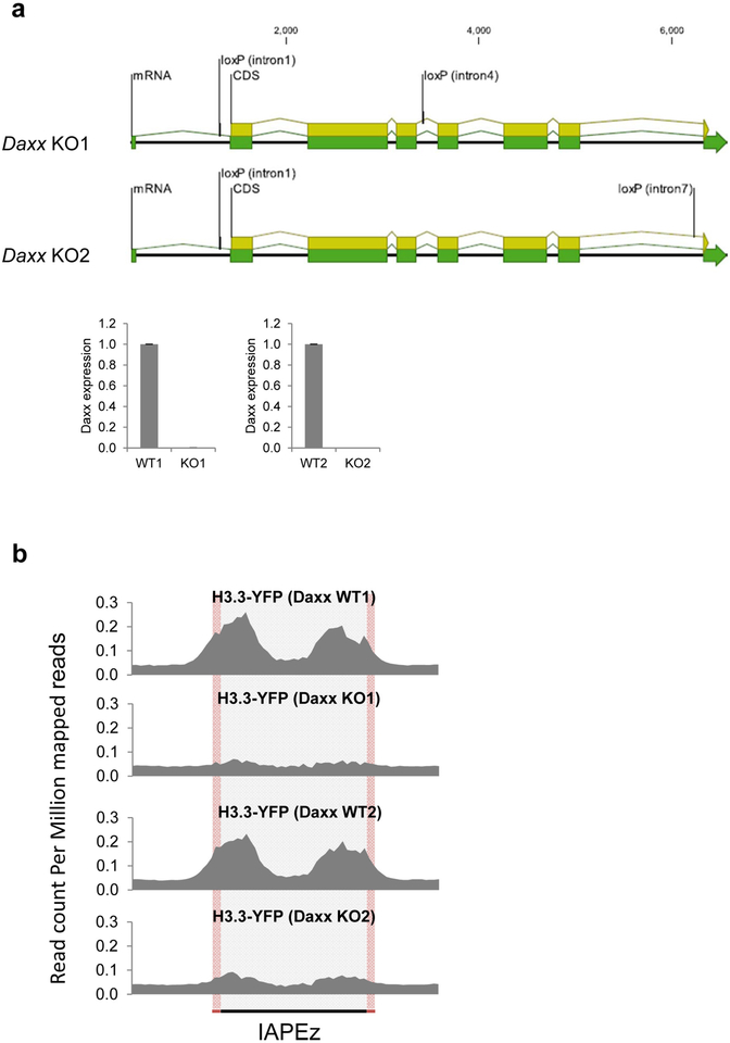
a, Top, representation of the two conditional Daxx-knockout alleles generated by CRISPR–Cas9-mediated loxP insertion at the indicated introns. Bottom, quantitative RT–PCR analysis of Daxx mRNA expression in Daxx-knockout ES cells. Data are mean ± s.d. expression (normalized to Gapdh) relative to the corresponding wildtype ES cells (n = 3, technical replicates). b, H3.3–YFP enrichment at 717 full-length IAP ERVs in two independently derived conditional Daxx-knockout ES cell lines.
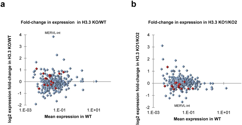
a, Mean fold change in ERV (LTR elements annotated in UCSC RepeatMasker) expression in H3.3-knockout ES cells (two cell lines) over wild-type ES cells (one cell line). b, Fold change in ERV expression comparing two H3.3-knockout ES cell lines. ERVs belonging to the IAP family are marked in red. Only ERV groups with more than 100 family members were considered for analysis.
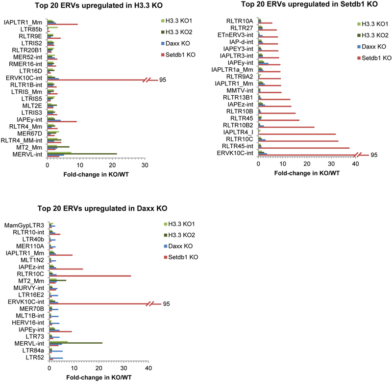
The fold change in expression over the corresponding wild-type control is shown for the top 20 upregulated ERV annotations in Setdb1-, H3.3- and Daxx-knockout ES cells. Annotations including –int represent the internal regions, which are transcribed from the cognate 5′ LTR, of the annotated ERV. For example, ERVK10C-int is the internal region of ERVK10C elements, with flanking LTRs: RLTR10A, RLTR10B and RLTR10C (depending on the specific genomic copy), which are also presented among the graphs. Similarly, IAPEz-int is the internal region of IAPEz elements with flanking cognate LTRs: IAPLTR1_Mm and IAPLTR1a_Mm, which are also represented. As the internal region is much longer and transcribed across its length, this is the most useful annotation to consider for expression analysis. The following published RNA-seq GEO data were re-analysed: GSM727424 (Setdb1-knockout ES cells); GSM1428580 and GSM1428581 (H3.3-knockout ES cells). Only ERV groups with more than 100 family members were considered for analysis.
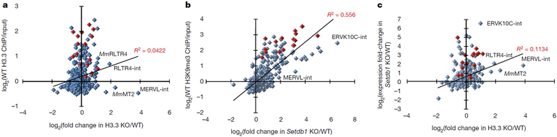
a, Mean fold change in ERV (long terminal repeat (LTR) elements annotated in UCSC RepeatMasker) expression in H3.3-knockout (KO) ES cells (two cell lines) over wild-type (WT) ES cells (one cell line) versus H3.3 enrichment in wild-type ES cells. ChIP, chromatin immunoprecipitation. b, Fold change in ERV expression in Setdb1-knockout ES cells over wild-type ES cells versus H3K9me3 enrichment in wild-type ES cells. c, Mean fold change in ERV expression in H3.3-knockout ES cells over wild-type ES cells versus fold change in ERV expression in Setdb1-knockout ES cells over wild-type ES cells. ERVs belonging to the IAP family are marked in red. Mm, mus musculus. Annotations including an ‘-int’ represent the internal regions, which are transcribed from the cognate 5′ LTR, of the annotated ERV. Only ERV groups with more than 100 family members were considered for analysis. Linear trend lines and corresponding R2 values are shown.
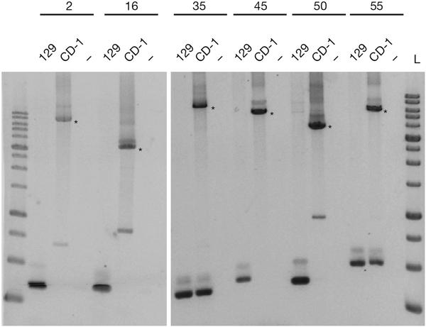
PCR assay of 129/SvJ and genomic DNA of the CD-1 feeders using primers flanking six ‘new’ copies described in H3.3-knockout ES cells in ref. . Asterisks indicate larger bands corresponding to the size expected for a typical IAP insertion. Non-template controls are shown (−) and 10-kb size ladders (L) are included on either side of the six copies. IAP copy indexes (2, 16, 35, 45, 50 and 55) are shown above the lanes and the coordinates can be found in Supplementary Table 1 along with the primer sequences.
Comment on
-
Histone H3.3 is required for endogenous retroviral element silencing in embryonic stem cells.Nature. 2015 Jun 11;522(7555):240-244. doi: 10.1038/nature14345. Epub 2015 May 4. Nature. 2015. PMID: 25938714 Free PMC article.
-
Elsässer et al. reply.Nature. 2017 Aug 2;548(7665):E7-E9. doi: 10.1038/nature23278. Nature. 2017. PMID: 28770850 No abstract available.
References
Publication types
Grants and funding
LinkOut - more resources
Full Text Sources
Other Literature Sources
Molecular Biology Databases
