

Pigment epithelium-derived factor promotes tumor metastasis through an interaction with laminin receptor in hepatocellular carcinomas
- PMID: 28771223
- PMCID: PMC5596550
- DOI: 10.1038/cddis.2017.359
Pigment epithelium-derived factor promotes tumor metastasis through an interaction with laminin receptor in hepatocellular carcinomas
Erratum in
-
Correction: Pigment epithelium-derived factor promotes tumor metastasis through an interaction with laminin receptor in hepatocellular carcinomas.Cell Death Dis. 2021 Jul 20;12(8):718. doi: 10.1038/s41419-021-03975-3. Cell Death Dis. 2021. PMID: 34285197 Free PMC article. No abstract available.
Abstract
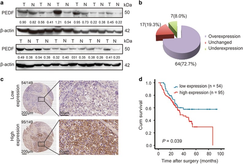
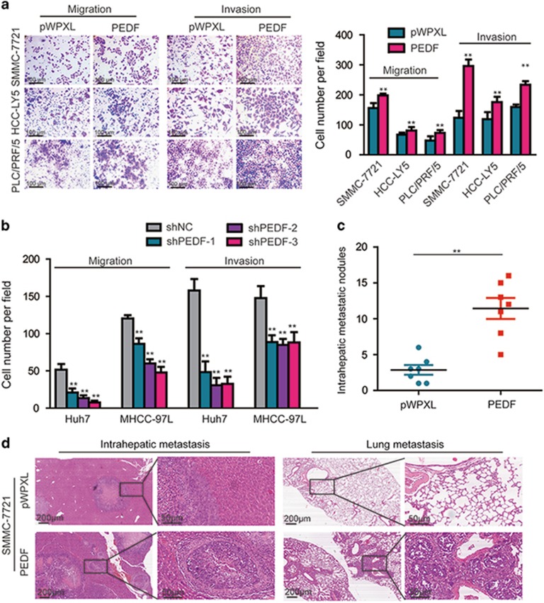
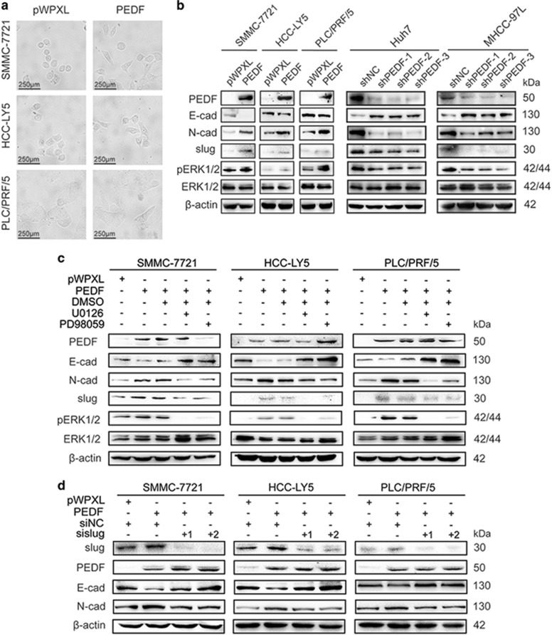
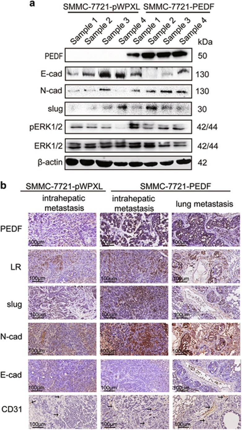
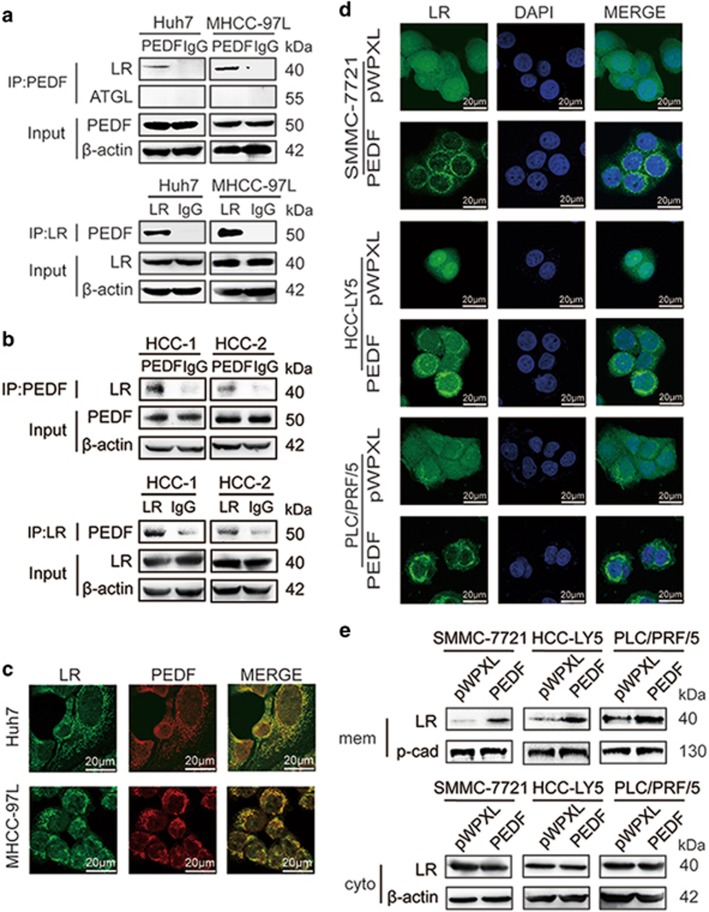
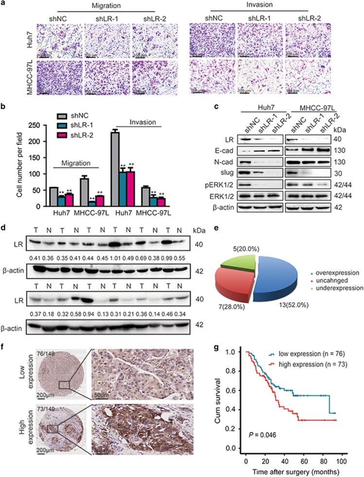
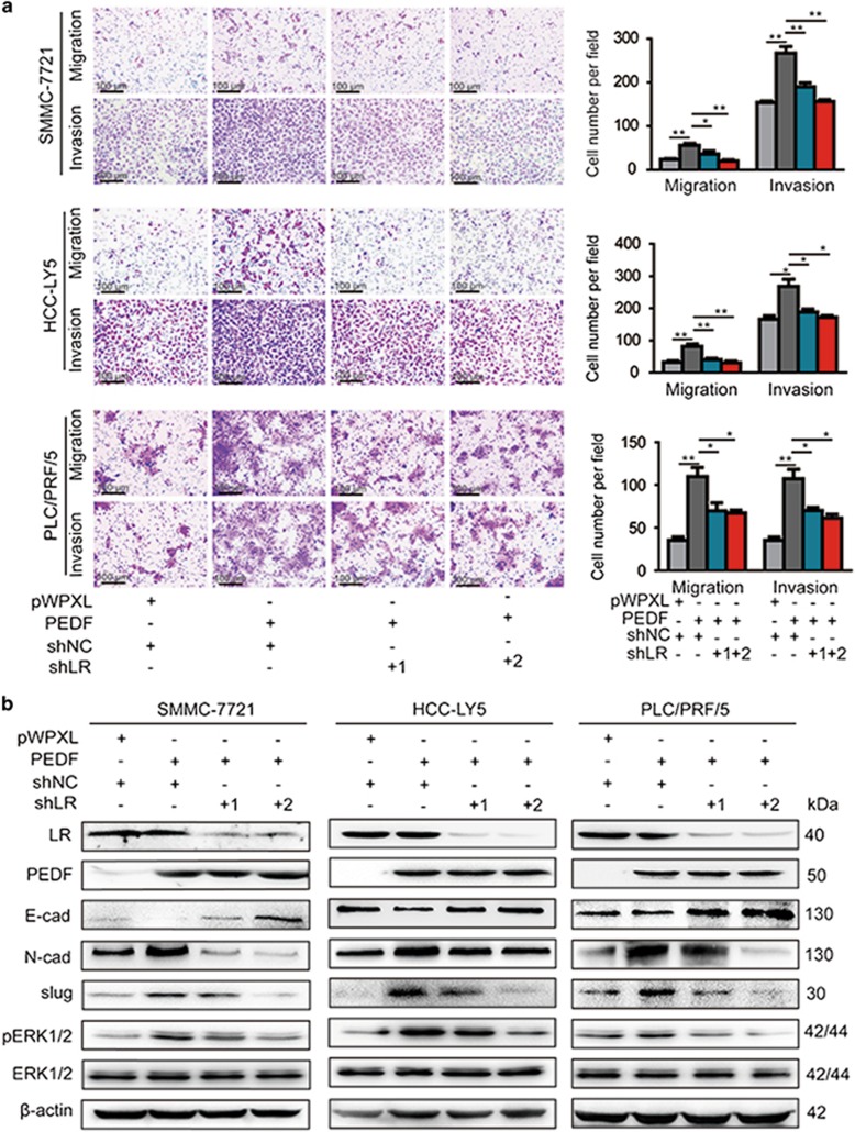
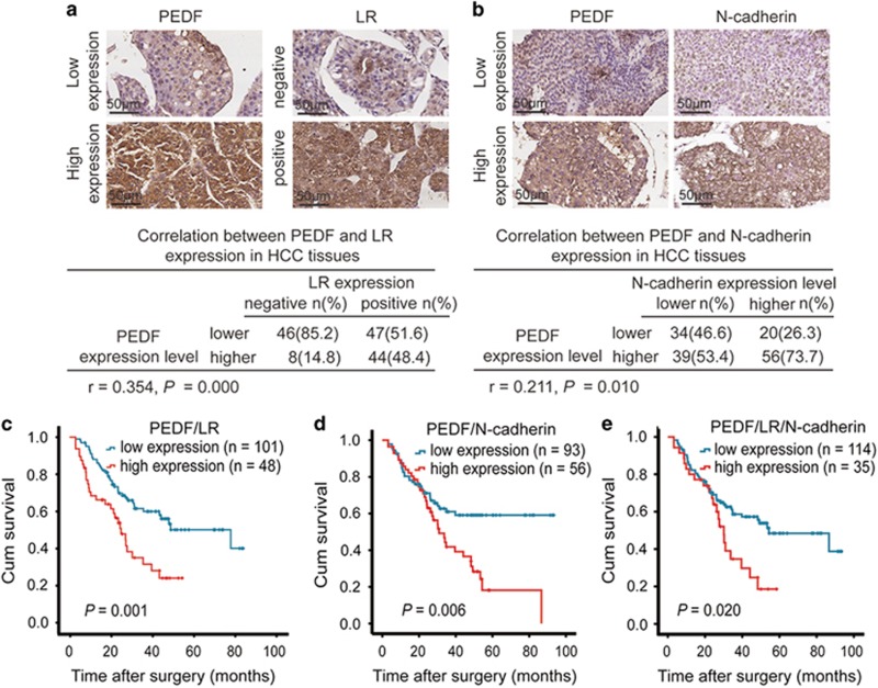
Pigment epithelium-derived factor (PEDF) has complex functions in tumor metastasis, but little is known about the roles of PEDF and its receptors in hepatocellular carcinoma (HCC). Here we found that high expression of PEDF is associated with shorter overall survival in HCC patients. Forced expression of PEDF enhanced HCC cell aggressive behavior in vitro and in vivo, whereas silencing PEDF expression reduced migration and invasion. Furthermore, PEDF expression led to changes in cell morphology and the expression of epithelial-mesenchymal transition (EMT)-related markers via ERK1/2 signaling pathway, including the upregulation of N-cadherin and slug, and the downregulation of E-cadherin in HCC cells. Our results further showed that PEDF could interact with laminin receptor (LR) and LR knockdown attenuated PEDF-induced migration, invasion and the change of EMT-related markers. More importantly, in clinical HCC specimens, we found that PEDF expression was correlated with subcellular localization of LR, and that high expression of PEDF and positive expression of LR predicted a poor prognosis. In conclusion, our results demonstrate a novel functional role of PEDF/LR axis in driving metastasis through ERK1/2-mediated EMT in HCC and provided a promising prognostic marker in HCC.
Conflict of interest statement
The authors declare no conflict of interest.
Figures
Similar articles
-
The contrary intracellular and extracellular functions of PEDF in HCC development.Cell Death Dis. 2019 Oct 3;10(10):742. doi: 10.1038/s41419-019-1976-4. Cell Death Dis. 2019. PMID: 31582735 Free PMC article.
-
Laminin receptor involvement in the anti-angiogenic activity of pigment epithelium-derived factor.J Biol Chem. 2009 Apr 17;284(16):10480-90. doi: 10.1074/jbc.M809259200. Epub 2009 Feb 17. J Biol Chem. 2009. PMID: 19224861 Free PMC article.
-
Pigment epithelium-derived factor inhibits lysosomal degradation of Bcl-xL and apoptosis in HepG2 cells.Am J Pathol. 2010 Jan;176(1):168-76. doi: 10.2353/ajpath.2010.090242. Epub 2009 Nov 30. Am J Pathol. 2010. PMID: 19948828 Free PMC article.
-
The increasing role of pigment epithelium-derived factor in metastasis: from biological importance to a promising target.Biochem Pharmacol. 2021 Nov;193:114787. doi: 10.1016/j.bcp.2021.114787. Epub 2021 Sep 24. Biochem Pharmacol. 2021. PMID: 34571004 Review.
-
Receptors that bind to PEDF and their therapeutic roles in retinal diseases.Front Endocrinol (Lausanne). 2023 Apr 17;14:1116136. doi: 10.3389/fendo.2023.1116136. eCollection 2023. Front Endocrinol (Lausanne). 2023. PMID: 37139333 Free PMC article. Review.
Cited by
-
Association of pigment epithelium derived factor expression with cancer progression and prognosis: a meta-analysis study.Discov Oncol. 2021 Dec 15;12(1):61. doi: 10.1007/s12672-021-00457-y. Discov Oncol. 2021. PMID: 35201465 Free PMC article.
-
Pedf derived peptides affect colorectal cancer cell lines resistance and tumour re-growth capacity.Oncotarget. 2019 Apr 26;10(31):2973-2986. doi: 10.18632/oncotarget.26085. eCollection 2019 Apr 26. Oncotarget. 2019. PMID: 31105879 Free PMC article.
-
Pigment epithelium-derived factor inhibits advanced glycation end product-induced proliferation, VEGF and MMP-9 expression in breast cancer cells via interaction with laminin receptor.Oncol Lett. 2021 Aug;22(2):629. doi: 10.3892/ol.2021.12890. Epub 2021 Jun 30. Oncol Lett. 2021. PMID: 34267821 Free PMC article.
-
Pigment Epithelium Derived Factor Is Involved in the Late Phase of Osteosarcoma Metastasis by Increasing Extravasation and Cell-Cell Adhesion.Front Oncol. 2022 Jan 31;12:818182. doi: 10.3389/fonc.2022.818182. eCollection 2022. Front Oncol. 2022. PMID: 35174090 Free PMC article.
-
Proteomic and Metabolomic Analysis of Bone Marrow and Plasma from Patients with Extramedullary Multiple Myeloma Identifies Distinct Protein and Metabolite Signatures.Cancers (Basel). 2023 Jul 25;15(15):3764. doi: 10.3390/cancers15153764. Cancers (Basel). 2023. PMID: 37568580 Free PMC article.
References
-
- Torre LA, Siegel RL, Ward EM, Jemal A. Global cancer incidence and mortality rates and trends—an update. Cancer Epidemiol Biomarkers Prev 2016; 25: 16–27. - PubMed
-
- Marquardt JU, Thorgeirsson SS. SnapShot: hepatocellular carcinoma. Cancer Cell 2014; 25: e551. - PubMed
-
- Becerra SP, Sagasti A, Spinella P, Notario V. Pigment epithelium-derived factor behaves like a noninhibitory serpin. Neurotrophic activity does not require the serpin reactive loop. J Biol Chem 1995; 270: 25992–25999. - PubMed
-
- Tombran-Tink J, Chader GG, Johnson LV. PEDF: a pigment epithelium-derived factor with potent neuronal differentiative activity. Exp Eye Res 1991; 53: 411–414. - PubMed
Publication types
MeSH terms
Substances
LinkOut - more resources
Full Text Sources
Other Literature Sources
Medical
Molecular Biology Databases
Research Materials
Miscellaneous
