. 2017 Aug 3;8(8):e2965.
doi: 10.1038/cddis.2017.353.

FGFR1 is critical for the anti-endothelial mesenchymal transition effect of N-acetyl-seryl-aspartyl-lysyl-proline via induction of the MAP4K4 pathway

Affiliations

FGFR1 is critical for the anti-endothelial mesenchymal transition effect of N-acetyl-seryl-aspartyl-lysyl-proline via induction of the MAP4K4 pathway

Jinpeng Li et al. Cell Death Dis. .

Abstract

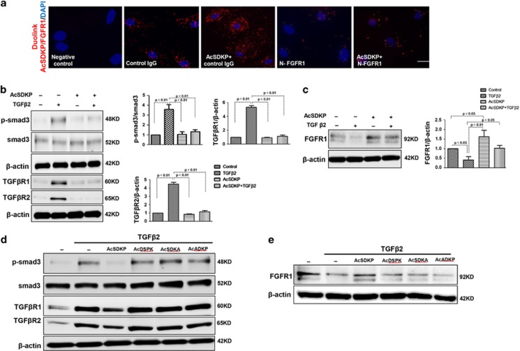
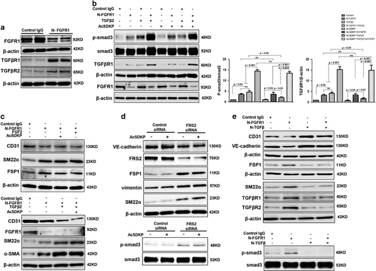
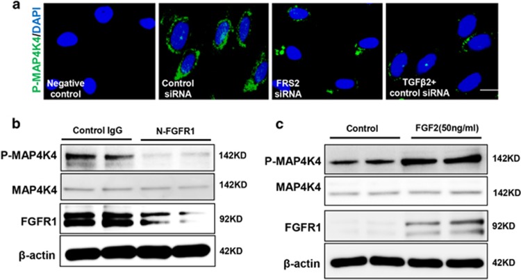
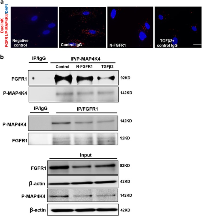
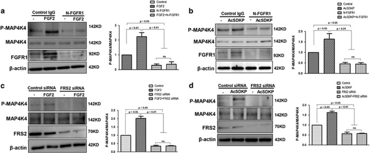
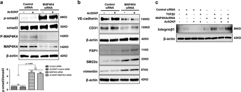
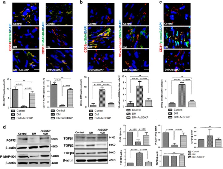
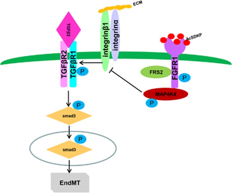
Endothelial-to-mesenchymal transition (EndMT) has been shown to contribute to organ fibrogenesis, and we have reported that the anti-EndMT effect of N-acetyl-seryl-aspartyl-lysyl-proline (AcSDKP) is associated with restoring expression of diabetes-suppressed fibroblast growth factor receptor (FGFR), the key anti-EndMT molecule. FGFR1 is the key inhibitor of EndMT via the suppression of the transforming growth factor β (TGFβ) signaling pathway, and mitogen-activated protein kinase kinase kinase kinase 4 (MAP4K4) inhibits integrin β1, a key factor in activating TGFβ signaling and EndMT. Here, we showed that the close proximity between AcSDKP and FGFR1 was essential for the suppression of TGFβ/smad signaling and EndMT associated with MAP4K4 phosphorylation (P-MAP4K4) in endothelial cells. In cultured human dermal microvascular endothelial cells (HMVECs), the anti-EndMT and anti-TGFβ/smad effects of AcSDKP were lost following treatment with a neutralizing FGFR1 antibody (N-FGFR1) or transfection of FRS2 siRNA. The physical interaction between FGFR1 and P-MAP4K4 in HMVECs was confirmed by proximity ligation analysis and an immunoprecipitation assay. AcSDKP induced P-MAP4K4 in HMVECs, which was significantly inhibited by treatment with either N-FGFR1 or FRS2 siRNA. Furthermore, MAP4K4 knockdown using specific siRNAs induced smad3 phosphorylation and EndMT in HMVECs, which was not suppressed by AcSDKP. Streptozotocin-induced diabetic CD-1 mice exhibited suppression of both FGFR1 and P-MAP4K4 expression levels associated with the induction of TGFβ/smad3 signaling and EndMT in their hearts and kidneys; those were restored by AcSDKP treatment. These data demonstrate that the AcSDKP-FGFR1-MAP4K4 axis has an important role in combating EndMT-associated fibrotic disorders.


Conflict of interest statement

The authors declare no conflict of interest.

Figures

Figure 1
Figure 1
Proximity between AcSDKP and FGFR1 inhibits the TGFβ/smad signaling pathway in HMVECs. (a) HMVECs were treated with N-FGFR1 (1.5  μg/ml) for 48 h with or without preincubation with AcSDKP (100 nM) for 2 h, and the proximity between AcSDKP and FGFR1 was analyzed by the Duolink In Situ Assay. For each slide, images at a × 400 original magnification were obtained from six different areas. (b and c) HMVECs were treated with TGFβ2 (5 ng/ml) for 15 min or 48 h with or without preincubation with AcSDKP for 2 h, and the p-smad3, TGFβR1, TGFβR2 and FGFR1 levels were analyzed by western blot. Densitometric analysis of the p-smad3/smad3, TGFβR1/β-actin, TGFβR2/β-actin and FGFR1/β-actin levels from each group (n=6) were analyzed. (d and e) HMVECs were incubated with TGFβ2 for 15 min or 48 h with or without preincubation with AcSDKP or its mutants (AcDSPK, AcSDKA, AcADKP) (100 nM) for 2 h. The p-smad3/smad3, TGFβR1/β-actin, TGFβR2/β-actin and FGFR1/β-actin protein levels were analyzed by western blot
Figure 2
Figure 2
AcSDKP suppresses TGFβ/smad signaling and EndMT through the FGFR1/FRS2 pathway. (a) HMVECs were treated with N-FGFR1 for 48 h, and the FGFR1, TGFβR1 and TGFβR2 protein levels were analyzed by western blot. (b) HMVECs were treated with TGFβ2 in the presence or absence of N-FGFR1 for 15 min with or without AcSDKP preincubation. The p-smad3 and TGFβR1 protein levels were analyzed by western blot. Densitometric analysis of the p-smad3/smad3 and TGFβR1/β-actin levels (n=3) in each group was performed. (c) HMVECs were incubated with either N-FGFR1 in the presence or absence of TGFβ2 for 48 h with or without preincubation with AcSDKP for 2 h or with N-FGFR1 in the presence or absence of TGFβ2 for 48 h with or without 24 h of incubation with FGF2 (50 ng/ml). The CD31, SM22α, FSP1 and α-SMA protein levels were analyzed by western blot. (d) HMVECs were transfected with FRS2 siRNA (100 nM) for 48 h with or without AcSDKP preincubation. The VE-cadherin, FSP1, vimentin, SM22α and p-smad3 levels were analyzed by western blot. (e) HMVECs were treated with N-FGFR1 for 48 h or 15 min in the presence or absence of N-TGFβ (1, 2, 3) (1.0  μg/ml). The CD31, VE-cadherin, SM22α, FSP1, TGFβR1, TGFβR2 and p-smad3 levels were analyzed by western blot
Figure 3
Figure 3
FGF2/FGFR1 mediates MAP4K4 signaling in endothelial cells. (a) Immunofluorescence microscopy analysis of P-MAP4K4 expression following FRS2 siRNA or TGFβ2 treatment. For each slide, images of six different fields of view at × 400 magnification were evaluated. The scale bar is 60 μm in each panel. (b and c) HMVECs were treated with N-FGFR1 for 48 h or FGF2 for 24 h. The P-MAP4K4 levels were analyzed by western blot
Figure 4
Figure 4
The proximity between FGFR1 and P-MAP4K4 decreases in FGFR1-deficient cells. (a) HMVECs were treated with N-FGFR1 or TGFβ2 for 48 h. The proximity between FGFR1 and P-MAP4K4 was analyzed using the Duolink In Situ Assay. For each slide, images at × 400 original magnification were obtained from six different areas. (b) immunoprecipitation analysis with either a P-MAP4K4 or a FGFR1 antibody was performed and analyzed by western blot. Then, the FGFR1 and P-MAP4K4 levels with N-FGFR1 or TGFβ2 treatment were analyzed by western blot in endothelial cells
Figure 5
Figure 5
MAP4K4 signaling is mediated by AcSDKP in a FGFR1/FRS2-dependent manner. (a and b) HMVECs were treated with N-FGFR1 for 48 h in the presence or absence of FGF2 or AcSDKP. P-MAP4K4 levels were analyzed by western blot. Densitometric analysis of P-MAP4K4 levels normalized to MAP4K4. For each group, n=3 were analyzed. (c and d) HMVECs were transfected with FRS2 siRNA for 48 h with or without FGF2 or AcSDKP treatment. P-MAP4K4 levels were analyzed by western blot. Densitometric analysis of P-MAP4K4 levels, normalized to MAP4K4. A total of n=3 from each group were analyzed
Figure 6
Figure 6
MAP4K4 deficiency induces TGFβ/smad signaling and EndMT via activation of integrin β1. (a) HMVECs were transfected with MAP4K4 siRNA (100 nM) for 48 h. Next, the cells were treated with or without AcSDKP for 2 h. The p-smad3/smad3 pathway was analyzed by western blot. Densitometric analysis of the p-smad3/smad3 levels was performed, with n=3 for each group. (b) HMVECs were treated with MAP4K4 siRNA for 48 h with or without AcSDKP treatment. The VE-cadherin, CD31, FSP1, SM22α and vimentin protein levels were analyzed by western blot. (c) HMVECs were transfected with MAP4K4 siRNA for 48 h in the presence or absence of TGFβ2 with or without AcSDKP. The integrin β1 level was analyzed by western blot
Figure 7
Figure 7
AcSDKP inhibits TGFβ/smad signaling and EndMT and restores the FGFR1 and P-MAP4K4 levels in diabetic hearts. (a) Immunofluorescence microscopy analysis of CD31/FGFR1 and CD31/P-MAP4K4 in the heart tissues from each group of mice. The scale bar is 60 μm in each panel. The CD31 and FGFR1 double-labeled cells and the CD31 and P-MAP4K4 double-labeled cells in each visual field were assessed by fluorescence microscopy and quantified. For each section, images from six different fields of view at × 400 magnification were evaluated. (b and c) Immunofluorescence microscopy analysis of CD31/α-SMA, VE-cadherin /SM22α and CD31/p-smad3 expression levels in the heart tissues from each group of mice. The scale bar is 60 μm in each panel. The CD31 and α-SMA double-labeled cells, the VE-cadherin and SM22α double-labeled cells and the CD31 and p-smad3 double-labeled cells in each visual field were analyzed by fluorescence microscopy and quantified. For each section, images from six different fields of view at × 400 magnification were evaluated. Four mice from each group were analyzed. (d) Western blot analysis of the FGFR1, P-MAP4K4, TGFβ1, TGFβ2 and TGFβ3 levels in cardiac tissues. A representative blot from four independent experiments was shown. The densitometric analysis of western blot data was presented (n=4). The diabetic mice are abbreviated as DM in the figure
Figure 8
Figure 8
Schematic of the AcSDKP/FGFR1/MAP4K4 pathway suppression of TGFβ/smad signaling and EndMT. In endothelial cells, the close proximity between AcSDKP and FGFR1 increased FGFR1 and induced its phosphorylation levels. Interacting with co-factor FRS2, FGFR1 recruited MAP4K4 and induced its phosphorylation. Subsequently, p-MAP4K4 suppressed integrinβ1 (integrinβ1 should be localized on the cell surface interacted with some of α integrins). Integrinβ1 was a potent activator of TGF-β signaling and also EndMT. Therefore, AcSDKP could inhibit EndMT through FGFR1-MAP4K4-dependent manner

Similar articles

Cited by

References

    1. Zeisberg EM, Potenta SE, Sugimoto H, Zeisberg M, Kalluri R. Fibroblasts in kidney fibrosis emerge via endothelial-to-mesenchymal transition. J Am Soc Nephrol 2008; 19: 2282–2287. - PMC - PubMed
    1. He J, Xu Y, Koya D, Kanasaki K. Role of the endothelial-to-mesenchymal transition in renal fibrosis of chronic kidney disease. Clin Exp Nephrol 2013; 17: 488–497. - PubMed
    1. Srivastava SP, Koya D, Kanasaki K. MicroRNAs in kidney fibrosis and diabetic nephropathy: roles on EMT and EndMT. Biomed Res Int 2013; 2013: 125469. - PMC - PubMed
    1. Zeisberg EM, Tarnavski O, Zeisberg M, Dorfman AL, McMullen JR, Gustafsson E et al. Endothelial-to-mesenchymal transition contributes to cardiac fibrosis. Nat Med 2007; 13: 952–961. - PubMed
    1. Potenta S, Zeisberg E, Kalluri R. The role of endothelial-to-mesenchymal transition in cancer progression. Br J Cancer 2008; 99: 1375–1379. - PMC - PubMed

MeSH terms

Substances

LinkOut - more resources