. 2017 Aug 3;8(8):e2966.
doi: 10.1038/cddis.2017.350.

Di (2-ethylhexyl) phthalate exposure impairs meiotic progression and DNA damage repair in fetal mouse oocytes in vitro

Affiliations

Di (2-ethylhexyl) phthalate exposure impairs meiotic progression and DNA damage repair in fetal mouse oocytes in vitro

Jing-Cai Liu et al. Cell Death Dis. .

Abstract

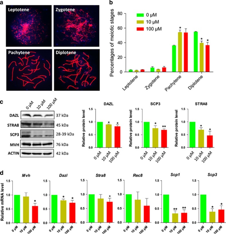
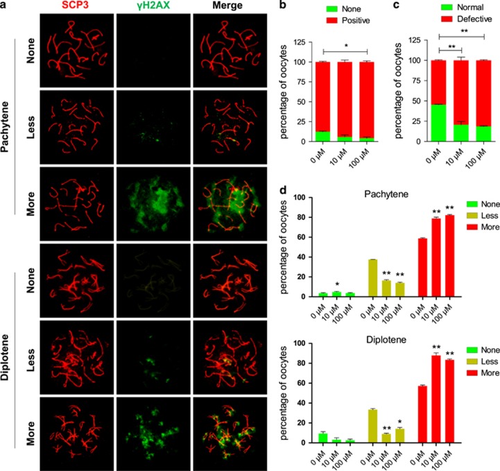
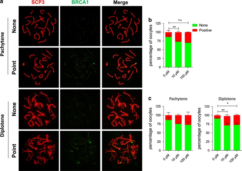
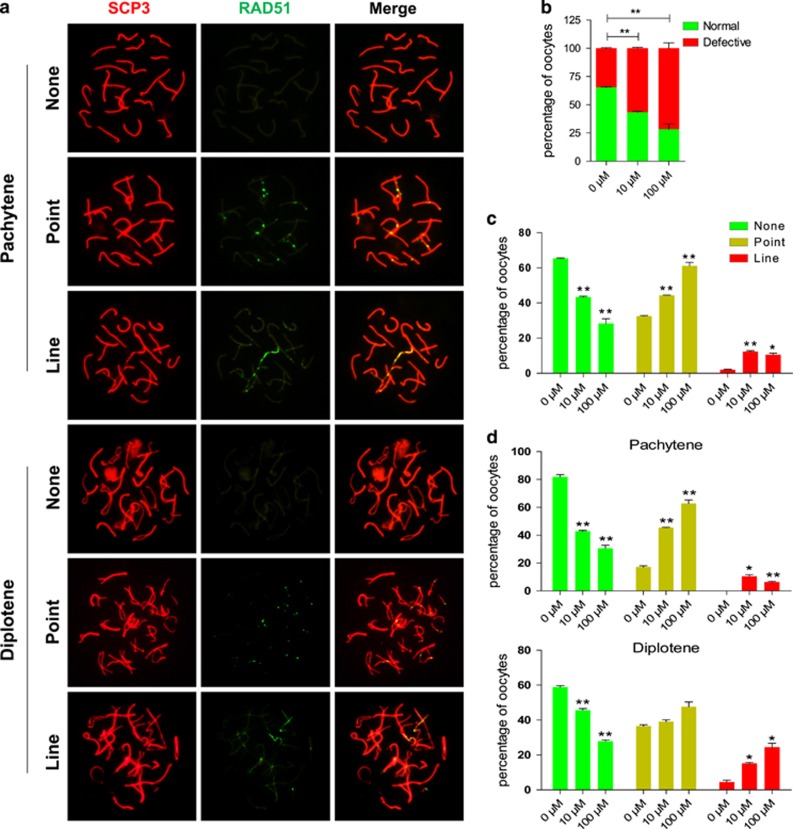
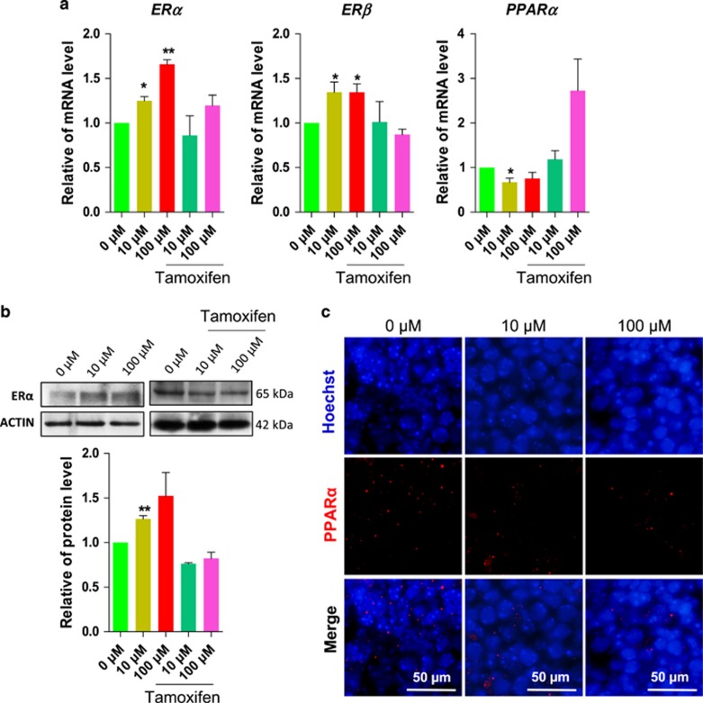
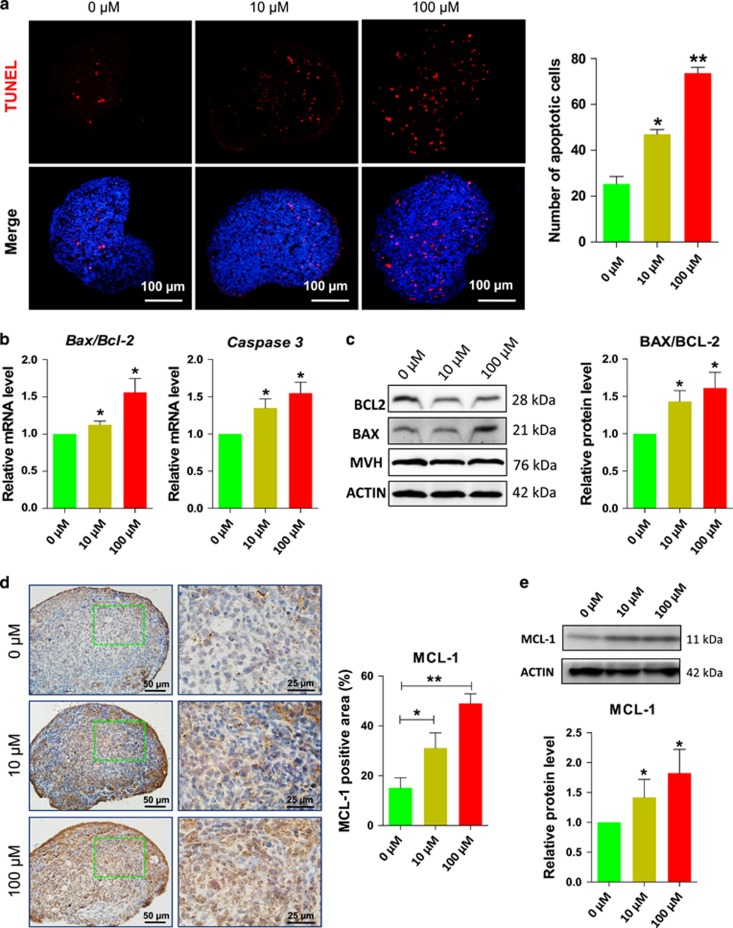
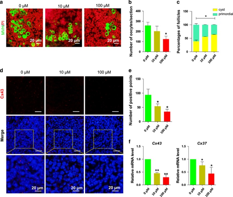
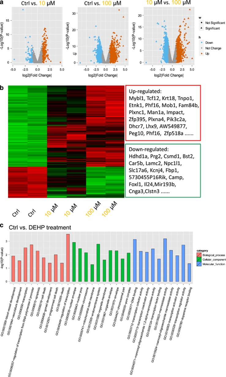
Di (2-ethylhexyl) phthalate (DEHP), is the most common member of the class of phthalates that are used as plasticizers and have become common environmental contaminants. A number of studies have shown that DEHP exposure impacts reproductive health in both male and female mammals by acting as an estrogen analog. Here, we investigated the effects of DEHP on meiotic progression of fetal mouse oocytes by using an in vitro model of ovarian tissue culture. The results showed that 10 or 100 μM DEHP exposure inhibited the progression of oocytes throughout meiotic prophase I, specifically from the pachytene to diplotene stages. DEHP possibly impairs the ability to repair DNA double-strand breaks induced by meiotic recombination and as a consequence activates a pachytene check point. At later stages, such defects led to an increased number of oocytes showing apoptotic markers (TUNEL staining, expression of pro-apoptotic genes), resulting in reduced oocyte survival, gap junctions, and follicle assembly in the ovarian tissues. Microarray analysis of ovarian tissues exposed to DEHP showed altered expression of several genes including some involved in apoptosis and gonad development. The expression changes of some genes clustered in cell-cell communication and signal transduction, along with plasma membrane, extracellular matrix and ion channel function classes, were dependent on the DEHP concentration. Together, these results bring new support to the notion that exposure to DEHP during gestation might exert deleterious effects on ovary development, perturbing germ cell meiosis and the expression of genes involved in a wide range of biological processes including ovary development.


Conflict of interest statement

The authors declare no conflict of interest.

Figures

Figure 1
Figure 1
Exposure to DEHP impairs meiotic progression of oocytes from pachytene to diplotene. (a) Immunolabeling of the oocyte chromosomes with anti-SYCP3 antibody (red) and Hoechst 33342 (blue). (b) Effect of DEHP on meiotic progression of oocytes throughout prophase I stages; percentage of each group is presented as mean±SD. Control: 36.27±0.80% pachytene and 54.99±0.66% diplotene; 10 μM and 100 μM DEHP 57.79±4.22% and 56.62±6.62% pachytene and 39.79±4.22% and 36.62±6.62% diplotene, respectively. (c) Representative WB showing the effect of DEHP on the expression of germ cell (DAZL) and meiotic (STRA8 and SCP3) specific proteins. (d) Effect of DEHP on the levels of mRNA in the ovarian tissues of germ cell (Mvh and Dazl), meiotic (Stra8, Rec8, Scp1 and Scp3). All experiments were repeated at least three times independently. (* P<0.05; ** P<0.01)
Figure 2
Figure 2
Effect of DEHP on γH2AX pattern. (a) Immunolabeling of the oocyte chromosomes with anti-SYCP3 (red) and anti-γH2AX (green) antibodies. (b) Percentage of positive γH2AX oocytes (79.36±3.97% and 81.40±0.62%, respectively), were significantly higher than that of control group (54.70±0.61% P<0.01). (c) Percentage of normal, negative or weak γH2AX staining and defective (strong γH2AX staining) oocytes after six days of culture with or without DEHP. (d) Percentage of oocytes at the pachytene and diplotene stages showing negative, weakly and strongly positive γH2AX staining after six days of culture with or without DEHP. (*P<0.05; **P<0.01)
Figure 3
Figure 3
Exposure to DEHP impairs DNA repair in oocytes. (a) Immunolabeling of the oocyte chromosomes with anti-SYCP3 (red) and anti-BRCA1 (green) antibodies. (b) Percentage of BRCA1 positive oocytes after six days of culture with or without DEHP (c) Percentage of oocytes at the pachytene and diplotene stages showing negative or positive BRCA1 staining after 6 days of culture with or without DEHP. (*P<0.05; **P<0.01)
Figure 4
Figure 4
Exposure to DEHP impairs DNA repair in oocytes. (a) Immunolabeling of the oocyte chromosomes with anti-SYCP3 (red) and anti-RAD51 (green) antibodies. (b) Percentage of oocytes showing none, RAD51 staining after six days of culture with or without DEHP. (c) Percentage of oocytes showing none, point or line RAD51 staining after six days of culture with or without DEHP. (d) Percentage of oocytes at the pachytene and diplotene stages showing none, point or line RAD51 staining after six days of culture with or without DEHP (*P<0.05; **P<0.01)
Figure 5
Figure 5
DEHP exposure affects the expression of ER and PPARα in the ovary. (a) Representative qRT-PCR for ERα, ERβ and PPARα transcripts in ovarian tissues cultured for 6 days in control, DEHP and DEHP plus tamoxifen. (b) Representative WB of ERα expression. (c) PPARα-staining of the ovarian tissues after 6 days of culture with or without DEHP. The percentage of each group is presented as mean±SD. All experiments were repeated at least three times. (*P<0.05; **P<0.01)
Figure 6
Figure 6
DEHP exposure promotes apoptosis in the ovary. (a) (Left) TUNEL-staining of the ovarian tissues after 6 days of culture with or without DEHP; (Right) Quantitative analyses of the number of TUNEL-positive cells. (b) Representative qRT-PCR for the apoptosis related genes Bax, Bcl-2 and Casp3 in ovarian tissues cultured for 6 days with or without DEHP. (c) (Left) representative WB of BAX and BCL-2; (Right) Quantitative analysis of BAX/BCL2 expression. (d) (Left) MCL-1-staining of the ovarian tissues after 6 days of culture with or without DEHP; (Right) Percentages of MCL-1 positive area. (e) (Up) Representative WB of MCL-1; (Down) Quantitative analysis of MCL-1 expression. The percentage of each group is presented as mean±SD. All experiments were repeated at least three times. (*P<0.05; **P<0.01)
Figure 7
Figure 7
DEHP exposure causes a reduction of oocyte number and of cyst breakdown (a) Control and DEHP exposed ovarian tissue stained for MVH (green specific for oocytes) after 10 days DEHP exposure, nuclei red; bar is 20 μM. (b) Number of oocytes in control and DEHP exposed groups. (c) Percentage of oocytes in cysts and primordial follicles in control and DEHP exposed groups. DEHP impairs the expression of connexins, (d) Control and DEHP exposed ovarian tissues stained for Cx43 (red), nuclei blue; bar is 20 μM. (e) Number of Cx43 positive gap junction plaques in control and DEHP exposed ovarian tissues. (f) qRT-PCR for Cx43 and Cx37. Relative fold changes are presented as mean±SD. All experiments were repeated at least three times. (* P<0.05; ** P<0.01)
Figure 8
Figure 8
Microarray analyses of gene expression in 12.5 dpc ovary cultured for 2 days in Control (Ctr) in the presence of DEHP. (a) Scatter plots of differentially expressed genes between Ctr and 10 μM DEHP, control and 100 μM DEHP, 10 μM and 100 μM DEHP groups. (b) Heatmap of differentially expressed genes between control and DEHP treated groups. (c) Gene ontology (GO) enrichment analysis of differentially expressed genes between Control (Ctr) and DEHP groups

References

    1. Lovekamp-Swan T, Davis BJ. Mechanisms of phthalate ester toxicity in the female reproductive system. Environ Health Perspect 2003; 111: 139–145. - PMC - PubMed
    1. Zhang XF, Zhang LJ, Li L, Feng YN, Chen B, Ma JM et al. Diethylhexyl phthalate exposure impairs follicular development and affects oocyte maturation in the mouse. Environ Mol Mutagen 2013; 54: 354–361. - PubMed
    1. Engel SM, Wolff MS. Causal inference considerations for endocrine disruptor research in children's health. Annu Rev Public Health 2013; 34: 139–158. - PMC - PubMed
    1. Lai FN, Liu JC, Li L, Ma JY, Liu XL, Liu YP et al. Di (2-ethylhexyl) phthalate impairs steroidogenesis in ovarian follicular cells of prepuberal mice. Arch Toxicol 2017; 91: 1279–1292. - PubMed
    1. Li L, Liu JC, Zhao Y, Lai FN, Yang F, Ge W et al. Impact of diethylhexyl phthalate on gene expression and development of mammary glands of pregnant mouse. Histochem Cell Biol 2015; 144: 389–402. - PubMed

Publication types

Substances

LinkOut - more resources