

Differential expression of pathogenic genes of Entamoeba histolytica vs E. dispar in a model of infection using human liver tissue explants
- PMID: 28771523
- PMCID: PMC5542602
- DOI: 10.1371/journal.pone.0181962
Differential expression of pathogenic genes of Entamoeba histolytica vs E. dispar in a model of infection using human liver tissue explants
Erratum in
-
Correction: Differential expression of pathogenic genes of Entamoeba histolytica vs E. dispar in a model of infection using human liver tissue explants.PLoS One. 2019 Jan 10;14(1):e0210895. doi: 10.1371/journal.pone.0210895. eCollection 2019. PLoS One. 2019. PMID: 30629702 Free PMC article.
Abstract
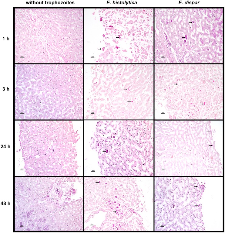
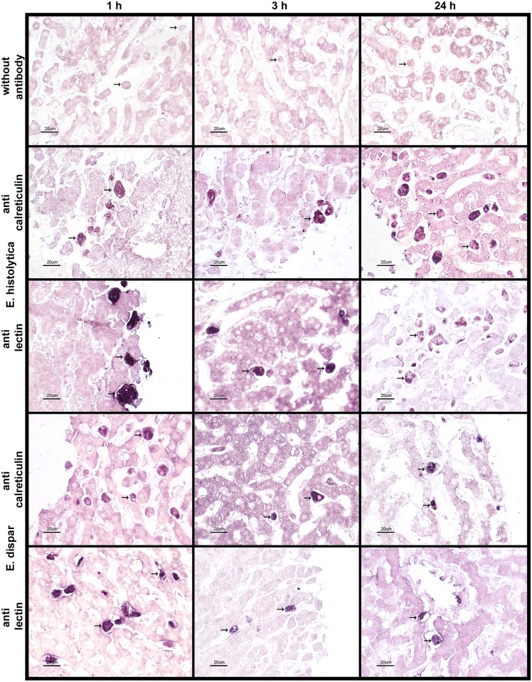
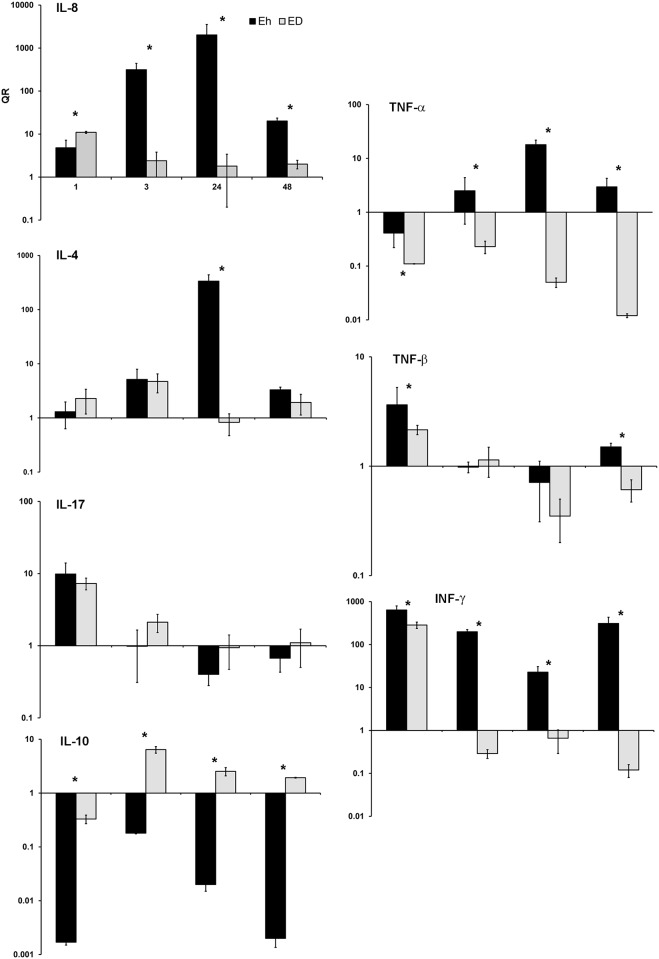
We sought to establish an ex vivo model for examining the interaction of E. histolytica with human tissue, using precision-cut liver slices (PCLS) from donated organs. E. histolytica- or E. dispar-infected PCLS were analyzed at different post-infection times (0, 1, 3, 24 and 48 h) to evaluate the relation between tissue damage and the expression of genes associated with three factors: a) parasite survival (peroxiredoxin, superoxide dismutase and 70 kDa heat shock protein), b) parasite virulence (EhGal/GalNAc lectin, amoebapore, cysteine proteases and calreticulin), and c) the host inflammatory response (various cytokines). Unlike E. dispar (non-pathogenic), E. histolytica produced some damage to the structure of hepatic parenchyma. Overall, greater expression of virulence genes existed in E. histolytica-infected versus E. dispar-infected tissue. Accordingly, there was an increased expression of EhGal/GalNAc lectin, Ehap-a and Ehcp-5, Ehcp-2, ehcp-1 genes with E. histolytica, and a decreased or lack of expression of Ehcp-2, and Ehap-a genes with E. dispar. E. histolytica-infected tissue also exhibited an elevated expression of genes linked to survival, principally peroxiredoxin, superoxide dismutase and Ehhsp-70. Moreover, E. histolytica-infected tissue showed an overexpression of some genes encoding for pro-inflammatory interleukins (ILs), such as il-8, ifn-γ and tnf-α. Contrarily, E. dispar-infected tissue displayed higher levels of il-10, the gene for the corresponding anti-inflammatory cytokine. Additionally, other genes were investigated that are important in the host-parasite relationship, including those encoding for the 20 kDa heat shock protein (HSP-20), the AIG-1 protein, and immune dominant variable surface antigen, as well as for proteins apparently involved in mechanisms for the protection of the trophozoites in different environments (e.g., thioredoxin-reductase, oxido-reductase, and 9 hypothetical proteins). Some of the hypothetical proteins evidenced interesting overexpression rates, however we should wait to their characterization. This finding suggest that the present model could be advantageous for exploring the complex interaction between trophozoites and hepatocytes during the development of ALA, particularly in the initial stages of infection.
Conflict of interest statement
Figures
Similar articles
-
Characteristics of inflammatory reactions during development of liver abscess in hamsters inoculated with Entamoeba nuttalli.PLoS Negl Trop Dis. 2018 Feb 8;12(2):e0006216. doi: 10.1371/journal.pntd.0006216. eCollection 2018 Feb. PLoS Negl Trop Dis. 2018. PMID: 29420539 Free PMC article.
-
Entamoeba histolytica: Overexpression of the gal/galnac lectin, ehcp2 and ehcp5 genes in an in vivo model of amebiasis.Parasitol Int. 2016 Dec;65(6 Pt A):665-667. doi: 10.1016/j.parint.2016.08.009. Epub 2016 Sep 5. Parasitol Int. 2016. PMID: 27616150
-
Host-Parasite interactions in Entamoeba histolytica and Entamoeba dispar: what have we learned from their genomes?Parasite Immunol. 2012 Feb-Mar;34(2-3):90-9. doi: 10.1111/j.1365-3024.2011.01325.x. Parasite Immunol. 2012. PMID: 21810102 Free PMC article. Review.
-
Differential expression of surface glycoconjugates on Entamoeba histolytica and Entamoeba dispar.Parasitol Int. 2009 Jun;58(2):171-7. doi: 10.1016/j.parint.2009.02.003. Epub 2009 Mar 6. Parasitol Int. 2009. PMID: 19269346
-
The pathogenicity of Entamoeba histolytica is related to the capacity of evading innate immunity.Parasite Immunol. 2005 Jan-Feb;27(1-2):1-8. doi: 10.1111/j.1365-3024.2005.00743.x. Parasite Immunol. 2005. PMID: 15813717 Review.
Cited by
-
Attenuation of In Vitro and In Vivo Virulence Is Associated with Repression of Gene Expression of AIG1 Gene in Entamoeba histolytica.Pathogens. 2023 Mar 21;12(3):489. doi: 10.3390/pathogens12030489. Pathogens. 2023. PMID: 36986411 Free PMC article.
-
The Interactions of Parasite Calreticulin With Initial Complement Components: Consequences in Immunity and Virulence.Front Immunol. 2020 Jul 23;11:1561. doi: 10.3389/fimmu.2020.01561. eCollection 2020. Front Immunol. 2020. PMID: 32793217 Free PMC article. Review.
-
Curcumin Attenuates the Pathogenicity of Entamoeba histolytica by Regulating the Expression of Virulence Factors in an Ex-Vivo Model Infection.Pathogens. 2019 Aug 15;8(3):127. doi: 10.3390/pathogens8030127. Pathogens. 2019. PMID: 31443160 Free PMC article.
-
A Novel Heat Shock Element (HSE) in Entamoeba histolytica that Regulates the Transcriptional Activation of the EhPgp5 Gene in the Presence of Emetine Drug.Front Cell Infect Microbiol. 2017 Nov 29;7:492. doi: 10.3389/fcimb.2017.00492. eCollection 2017. Front Cell Infect Microbiol. 2017. PMID: 29238701 Free PMC article.
-
The Dichloromethane Fraction of Croton sonorae, A Plant Used in Sonoran Traditional Medicine, Affect Entamoeba histolytica Erythrophagocytosis and Gene Expression.Front Cell Infect Microbiol. 2021 Jul 23;11:693449. doi: 10.3389/fcimb.2021.693449. eCollection 2021. Front Cell Infect Microbiol. 2021. PMID: 34368014 Free PMC article.
References
-
- Lohia A. The cell cycle of Entamoeba histolytica. Mol Cell Biochem. 2003; 253: 217–222. - PubMed
-
- Sargeaunt PG, Williams JE, Grene JD. The differentiation of invasive and noninvasive Entamoeba histolytica by isoenzyme electrophoresis. Trans R Soc Trop Med Hyg. 1978; 72: 519–521. - PubMed
-
- Diamond LS, Clark CG. A redescription of Entamoeba histolytica Schaudinn, 1903 (Emended Walker, 1911) separating it from Entamoeba dispar Brumpt, 1925. J Eukaryot Microbiol. 1993; 40:340–344. - PubMed
-
- WHO/PAN American Health Organization/UNESCO. Expert Consultation on Amoebiasis. WHO Weekly Epidem Rec 1997; 72: 97–100
MeSH terms
Substances
LinkOut - more resources
Full Text Sources
Other Literature Sources
Research Materials
