. 2017 Aug 3;12(8):e0182427.
doi: 10.1371/journal.pone.0182427. eCollection 2017.

Human leucocyte antigen (HLA-DR) gene expression is reduced in sepsis and correlates with impaired TNFα response: A diagnostic tool for immunosuppression?

Affiliations

Human leucocyte antigen (HLA-DR) gene expression is reduced in sepsis and correlates with impaired TNFα response: A diagnostic tool for immunosuppression?

Martin Sebastian Winkler et al. PLoS One. .

Abstract

Background: Sepsis is defined as a dysregulated immune response to infection. Impaired immune response in sepsis, often described as endotoxin tolerance, is characterized by unresponsiveness of monocytes on lipopolysaccharide (LPS) stimulation to release tumor necrosis factor α (TNFα). Furthermore, decreased monocyte surface protein expression of human leucocyte antigen DR (HLA-DR) is a marker for changes of the innate immune response during sepsis. Quantitative polymerase chain reaction (qPCR) and flow-cytometry (FACS) have been used to measure protein or gene expression of HLA-DR. We aimed to determine whether changes in mRNA expression of HLA-DR are associated with impaired TNFα response in human sepsis.

Methods: Surface protein together with mRNA expression of HLA-DR were measured by FACS and qPCR in a cohort of 9 sepsis patients and compared to 10 pre-operative control patients in a prospective study. In addition, 20 patients with post-surgical inflammation, 20 patients with sepsis or septic shock were included and TNFα was determined following ex vivo stimulation of whole blood with 500 pg/mL LPS. Total RNA was prepared from whole blood and subjected to qPCR analysis for expression analysis of HLA-DR alpha (HLA-DRA) to correlate TNFα response with HLA-DRA expression.

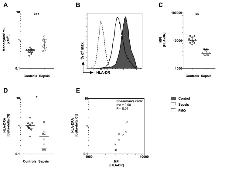
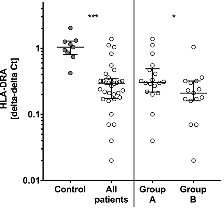
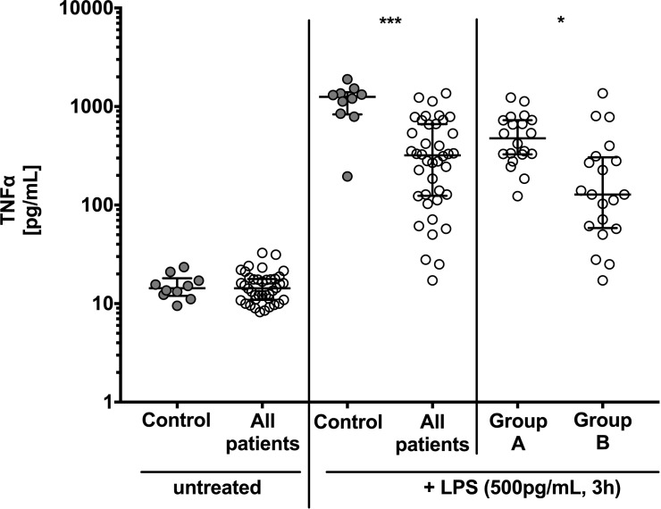
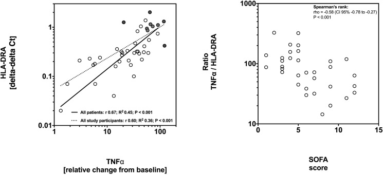
Results: Patients with sepsis presented higher numbers of monocytes in peripheral blood (P<0.001) but decreased surface protein and mRNA HLA-DR levels when compared to controls. In all patients mRNA expression of HLA-DRA was decreased by approximately 70% compared to controls (P<0.01) and was lowest in patients with sepsis or septic shock (P<0.01). TNFα response to LPS was decreased in all patients (median 319 pg/mL versus controls 1256 pg/mL; P<0.01) and lowest in patients with sepsis or septic shock (median 128 pg/mL; P<0.01). HLA-DRA correlated positively with TNFα response in all study participants (r +0.60, P<0.001) and within patients (r +0.67, P<0.001). The TNFα:HLA-DRA ratio correlated negatively with severity and the Sequential Organ Failure Assessment (SOFA) score (Spearman's rho -0.59, P<0.001).

Conclusion: In this study, HLA-DRA expression was associated with a functional assay of the innate immune response. Future interventional studies aimed at the immune response during sepsis could make use of these methods for optimizing target groups based on biological plausibility and intervention effectiveness.


Conflict of interest statement

Competing Interests: The authors have declared that no competing interests exist.

Figures

Fig 1
Fig 1. Monocytosis but loss of protein surface and gene expression of human leucocyte antigen DR (HLA-DR) is observed in sepsis patients.
(A) Number of monocytes in cells/mL shows significant higher numbers of monocytes/mL in sepsis patients (n = 9) compared to controls (n = 10). (B) Protein surface expression levels of HLA-DR by monocytes are shown as a representative histogram and (C) with the corresponding analysis of median fluorescence intensity (MFI). Sepsis patients show reduced HLA-DR surface expression compared to controls. (A-C) Whole blood was stained for cell surface molecules (CD14 and HLA-DR) and analyzed by flow cytometry (FACS). Data are presented as box and whisker plots with median and interquartile range and statistical analysis was performed using non-parametric Mann-Whitney-U test. (D) Gene expression of HLA-DRA is reduced in sepsis patients. mRNA was prepared from whole blood and HLA-DRA expression levels were assessed by quantitative PCR (qPCR). HLA-DRA expression levels are normalized to internal control gene peptidylpropylisomerase B (PPIB). Data are presented using the delta-delta Ct method and as box and whisker plots showing the median with interquartile range. Statistical analysis was performed using non-parametric Mann-Whitney-U test. (E) Spearman’s rank correlation is presented with median fluorescence intensity (MFI) for HLA-DR protein on the x-axis and mRNA expression of HLA-DR presented as delta-delta Ct on the y-axis. Each circle represents a data set from an individual patient. CD: cluster of differentiation, mRNA: messenger RNA, FMO: Fluorescence Minus One controls, PCR: polymerase chain reaction. P<0.05:*; P<0.01:**; P<0.001:***.
Fig 2
Fig 2. mRNA expression of human leucocyte antigen DR alpha (HLA-DRA) is lowest in sepsis patients.
HLA-DRA levels are significant reduced in all patients (n = 40) when compared to controls (n = 10). HLA-DRA levels in patients with sepsis (group B, n = 10) are lowest and significantly reduced when compared to postoperative patients (group A, n = 10). Total mRNA was prepared from whole blood and HLA-DRA expression were assessed by quantitative polymerase chain reaction (qPCR). mRNA levels are normalized to peptidylpropylisomerase B (PPIB) using the delta-delta Ct method. Box and whisker plots with median and interquartile range are presented and statistical analysis was performed using non-parametric Mann-Whitney-U test. P<0.05:*; P<0.001:***.
Fig 3
Fig 3. Tumor necrosis factor α (TNFα) response to lipopolysaccharide (LPS) is lowest in sepsis patients.
There is no difference in baseline TNFα levels of untreated whole blood in controls and patients. When stimulated with LPS TNFα responsiveness is significant reduced in all patients compared to controls and is lower in sepsis patients (group B) compared to patients after surgery (group A). TNFα levels were determined in the supernatant of untreated whole blood and after stimulation with 500 pg/mL LPS for 3h in controls (n = 10), patients with post-surgical inflammation (group A; n = 20) and patients with sepsis and septic shock (group B, n = 20). Data are presented as box and whisker plots with median and interquartile range. Statistical analysis was performed using non-parametric Mann-Whitney-U test. P<0.05: *, P<0.001: ***.
Fig 4
Fig 4. Low expression of human leucocyte antigen receptor alpha (HLA-DRA) is associated with unresponsiveness of whole blood to lipopolysaccharide (LPS) and is associated with disease severity.
(A) Whole blood was stimulated with 500 pg/mL LPS for 3h and TNFα levels were determined in the supernatant. The x-axis shows fold changes from baseline TNFα levels after ex vivo stimulation. The y-axis shows HLA-DRA expression levels assessed by quantitative polymerase chain reaction (qPCR) using the delta-delta Ct method. Linear regression analysis was performed after transformation to a logarithmic scale to reach normal distribution. Solid line: regression for patients; dotted line: regression for all study participants. Circles represent data from patients and dots represent data from controls. (B) TNFα:HLA-DRA ratio correlates with the SOFA score. Low TNFα response to LPS stimulation is associated with low expression levels of HLA-DRA as shown in (A). The ratio TNFα:HLA-DRA correlates with severity of the disease as measured by the SOFA score. Spearman’s rank test was used to calculate correlation. Circles represent ratios from individual patients.

References

    1. Deutschman CS, Tracey KJ. Sepsis: current dogma and new perspectives. Immunity. 2014;40(4):463–75. doi: 10.1016/j.immuni.2014.04.001 . - DOI - PubMed
    1. Hamers L, Kox M, Pickkers P. Sepsis-induced immunoparalysis: mechanisms, markers, and treatment options. Minerva anestesiologica. 2015;81(4):426–39. . - PubMed
    1. Boomer JS, Green JM, Hotchkiss RS. The changing immune system in sepsis: Is individualized immuno-modulatory therapy the answer? Virulence. 2013;5(1). Epub 2013/09/27. doi: 10.4161/viru.26516 - DOI - PMC - PubMed
    1. Wiedermann CJ. Adjuvant treatment of sepsis: what is known? Medizinische Klinik, Intensivmedizin und Notfallmedizin. 2014;109(8):583–90. doi: 10.1007/s00063-014-0379-7 . - DOI - PubMed
    1. Hou T, Huang D, Zeng R, Ye Z, Zhang Y. Accuracy of serum interleukin (IL)-6 in sepsis diagnosis: a systematic review and meta-analysis. Int J Clin Exp Med. 2015;8(9):15238–45. ; PubMed Central PMCID: PMCPMC4658898. - PMC - PubMed

MeSH terms