. 2017 Aug 3;12(8):e0181806.
doi: 10.1371/journal.pone.0181806. eCollection 2017.

IL-25-induced activation of nasal fibroblast and its association with the remodeling of chronic rhinosinusitis with nasal polyposis

Affiliations

IL-25-induced activation of nasal fibroblast and its association with the remodeling of chronic rhinosinusitis with nasal polyposis

Soo-Kyoung Park et al. PLoS One. .

Abstract

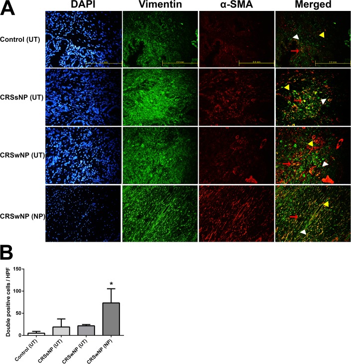
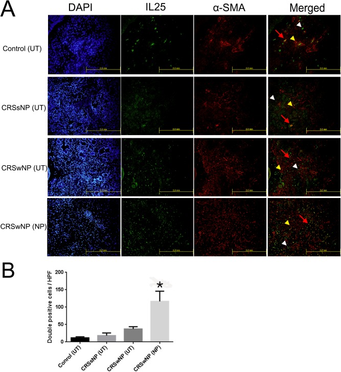
Background and objective: Interleukin (IL)-25 has been shown to play an important role in the pathogenesis of chronic rhinosinusitis with nasal polyps. Nasal polyps are associated with chronic inflammation of the mucous membranes in the paranasal sinuses and are involved in extracellular matrix (ECM) accumulation. The aim of this study is to evaluate the effects of IL-25 on myofibroblast differentiation, ECM production and the expression of matrix metalloproteinases in nasal polyp derived fibroblasts (NPDFs) and to determine the molecular mechanism underlying these processes.

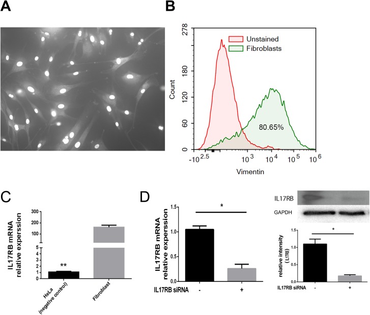
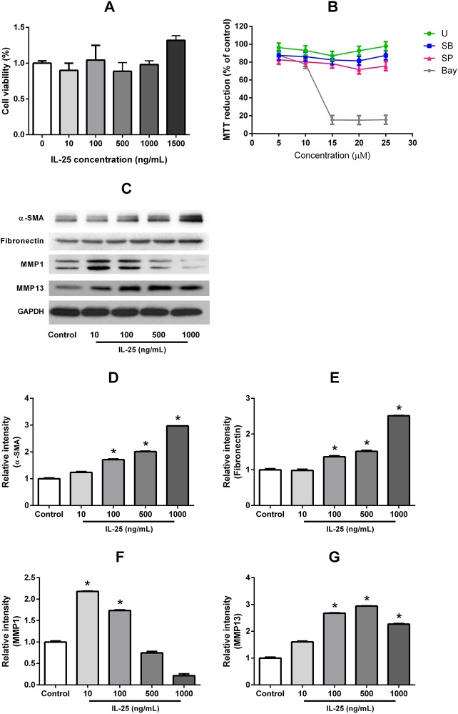
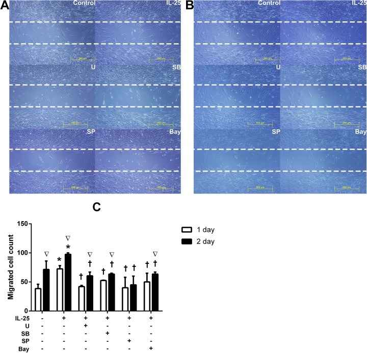
Materials and methods: A total of 40 patients were enrolled in this study for Immunofluorescence studies. Expression of IL17 receptor B was evaluated by real time reverse transcription polymerase chain reaction (PCR) in NPDFs. NPDFs were stimulated with IL-25 for 48 h in the presence or absence of mitogen-activated protein kinase (MAPK) and NF-κB inhibitors or small interfering RNAs (siRNA). The protein levels of fibrosis active mediators were examined using western blotting. Fibroblast migration was evaluated with a scratch assay. The total collagen amount was analyzed with the Sircol collagen assay.

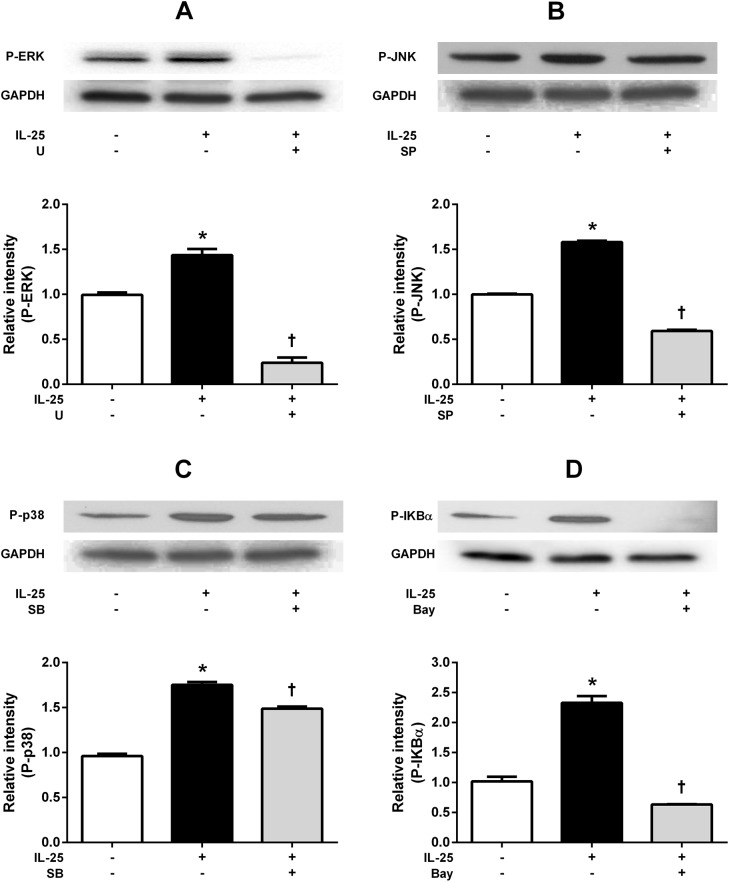
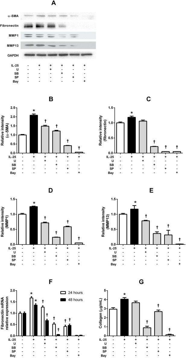
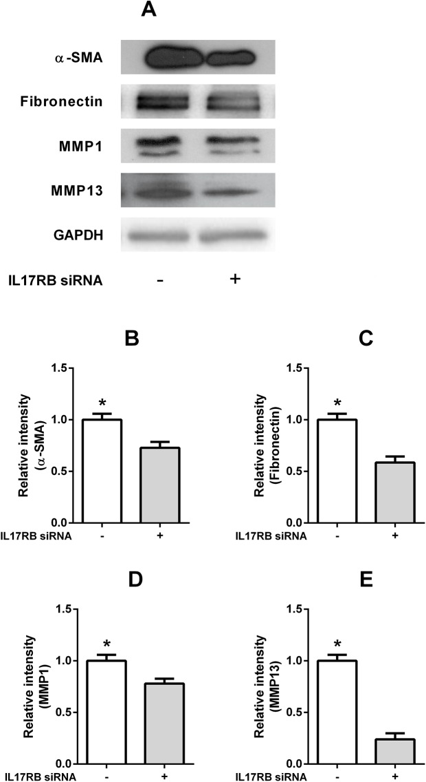
Results: IL-25 induced α-SMA, fibronectin, and MMP-1 and -13, which were dependent on IL-17RB. IL-25 also induced activation of NF-κB and mitogen-activated protein kinase (MAPKs). By using the specific inhibitor of ERK, p38, JNK and NF-κB (U, SB, SP and Bay), we found that IL-25-induced expressions of α-SMA, fibronectin, and MMPs was regulated by the signaling pathways of MAPKs and NF-κB. IL-25 also induces α-SMA, fibronectin, and MMPs expression through IL-17RB-dependent pathways in NPDFs. The increased migration ability induced by IL-25 was suppressed by the specific inhibitors of MAPKs and NF-κB.

Conclusion: Our data indicate that IL-25 induced myofibroblast differentiation, fibronectin production, and MMP-1 and -13 expressions through the signaling pathways of MAPKs and NF-κB. in NPDFs and increased expression of IL-25 were also involved in the pathogenesis of nasal polyposis by affecting nasal fibroblasts in chronic rhinosinusitis with nasal polyps.


Conflict of interest statement

Competing Interests: The authors have declared that no competing interests exist.

Figures

Fig 1
Fig 1. Double–immunofluorescent staining of myofibroblasts in the tissues of the four groups.
Double-immunofluorescence staining was undertaken to colocalize the cells with Vimentin (green color)/ α-SMA (red color) among the groups (A). The number of double positive cells (Vimentin+ α-SMA+) was considerably higher in the NP tissues of the CRSwNP group compared to the other groups (*p < 0.05) (B). Vimentin: white arrow head, α-SMA: yellow arrow head, double positive cells: red arrow. Triple tests were performed on all of the experiments.
Fig 2
Fig 2. Double–immunofluorescent staining of the correlation between IL-25 level and myofibroblast in the tissues of the four groups.
Double-immunofluorescence staining was undertaken to colocalize the cells with IL-25 (green color)/ α-SMA (red color) among the groups (A). The number of double positive cells (IL-25+ α-SMA+) was considerably higher in the NP tissues of CRSwNP group compared to the other groups. Vimentin and IL-25: white arrow head, α-SMA: yellow arrow head, double positive cells: red arrow (*p < 0.05). Triple tests were performed on all of the experiments.
Fig 3
Fig 3. Microscopic morphology of NPDFs and expression of IL-25 receptor on NPDFs.
Microscopic morphology of NPDFs (A). Approximately 80% of the cells in the cultured NPDFs were positive for vimentin (B). The mRNA expression of IL-17 receptor B (IL-17RB) was evaluated using RT-PCR (C), the IL-17RB mRNA and IL-17RB protein expression levels were significantly suppressed by IL-17RB siRNA compared with the control siRNA (*p < 0.05) (D). Triple tests were performed on all of the experiments.
Fig 4
Fig 4. IL-25 induced myofibroblast differentiation (α-SMA), fibronectin production and MMPs expression in NPDFs.
An MTT assay was performed to exclude the possibility of IL-25-induced and specific inhibitors-induced cellular cytotoxicity of the NPDFs (A). The specific inhibitors did not affect cell viability (B) The expression levels of α-SMA, fibronectin, MMP-1 and MMP-13 protein were determined by western blot assays (*p < 0.05) (C-G). Triple tests were performed on all of the experiments.
Fig 5
Fig 5. IL-25 induced activation of MAPKs and NF-κB in NPDF.
Effects of IL-25 on the activation of mitogen-activated protein kinases (ERK, p38, JNK) and NF-κB evaluated by Western blotting. Phosphorylation of representative MAPKs, ERK (A), JNK (B), and p38 MAPK (C) was induced by IL-25 treatment, after which they became inhibited by their specific inhibitors, including U, SP, and SB, respectively. (p < 0.05). IL-25 induced phosphorylation of IκBα; p-IκBα expression was inhibited by Bay (p < 0.05) (D). Values are expressed as means ± standard errors of independent experiments. (*p < 0.05 vs control). †p <0.05 vs IL-25 alone. Triple tests were performed on all of the experiments. U = specific inhibitor of ERK, SB = specific inhibitor of p38, SP = specific inhibitor of JNK, Bay = specific inhibitor of U = specific inhibitor of NF-κB.
Fig 6
Fig 6
Regulation of IL-25-induced α-SMA, fibronectin and MMPs expression through various signaling pathways in NPDFs. (A) The expression levels of α-SMA, fibronectin, MMP-1, and MMP-13 were determined by western blot assay or RT-PCR. Treatments with the specific inhibitors significantly inhibited IL-25-induced α-SMA (B), MMP-1 (D), and MMP-13 (E) protein expression (p < 0.05). IL-25-induced fibronectin (C) expression was inhibited by pretreatment with NF-κB, JNK, and p38 pathway inhibitors, but not by the ERK inhibitor. IL-25-induced fibronectin mRNA expression level was measured by RT- PCR (F). The total soluble collagen level was measured by collagen assay (G) Values are expressed as means ± standard errors of independent experiments. *P<0.05 vs control; †p <0.05 vs IL-25 alone. Triple tests were performed on all of the experiments. U = specific inhibitor of ERK, SB = specific inhibitor of p38, SP = specific inhibitor of JNK, Bay = specific inhibitor of U = specific inhibitor of NF-κB.
Fig 7
Fig 7. IL-25 induced α-SMA, fibronectin and MMPs expressions via IL-17RB dependent pathways in NPDFs.
Knock down of IL-17RB by IL-17RB siRNA decreased IL-25-induced a-SMA, fibronectin and MMPs expression in NPDFs (A, B, C, D, E). Values are expressed as means ± standard errors of independent experiments. *P<0.05 vs control. Triple tests were performed on all of the experiments.
Fig 8
Fig 8. Migration of NPDFs facilitated by IL-25 facilitated the migration of NPDFs.
Migration cells were examined by a cell migration assay. Photos of our microscopic observations of cell migration assays showing nasal fibroblast migration by IL-25 and pretreatment with specific inhibitors after 24h (A) and 48h (BC). The number of fibroblasts was counted in a ‘cell-free gap’ (500μm in width) after 24h and 48h (C). Values are expressed as means ± standard errors of independent experiments. *P<0.05 vs control; †p <0.05 vs IL-25 alone; ∇p <0.05 vs 24hr. Scale bar = 500 μm; Triple tests were performed on all of the experiments. U = specific inhibitor of ERK, SB = specific inhibitor of p38, SP = specific inhibitor of JNK, Bay = specific inhibitor of U = specific inhibitor of NF-κB.

Similar articles

Cited by

References

    1. Fokkens WJ, Lund VJ, Mullol J, Bachert C, Alobid I, Baroody F, et al. European Position Paper on Rhinosinusitis and Nasal Polyps 2012. Rhinology Supplement. 2012;(23):3 p preceding table of contents, 1–298. Epub 2012/07/07. . - PubMed
    1. Redington AE. Fibrosis and airway remodelling. Clinical and experimental allergy: journal of the British Society for Allergy and Clinical Immunology. 2000;30 Suppl 1:42–5. Epub 2000/06/13. . - PubMed
    1. Xu J, Han R, Kim DW, Mo JH, Jin Y, Rha KS, et al. Role of Interleukin-10 on Nasal Polypogenesis in Patients with Chronic Rhinosinusitis with Nasal Polyps. PloS one. 2016;11(9):e0161013 Epub 2016/09/02. doi: 10.1371/journal.pone.0161013 ; PubMed Central PMCID: PMCPMC5008817. - DOI - PMC - PubMed
    1. Gabasa M, Royo D, Molina-Molina M, Roca-Ferrer J, Pujols L, Picado C, et al. Lung myofibroblasts are characterized by down-regulated cyclooxygenase-2 and its main metabolite, prostaglandin E2. PloS one. 2013;8(6):e65445 Epub 2013/06/12. doi: 10.1371/journal.pone.0065445 ; PubMed Central PMCID: PMCPMC3670886. - DOI - PMC - PubMed
    1. Watelet JB, Van Zele T, Gjomarkaj M, Canonica GW, Dahlen SE, Fokkens W, et al. Tissue remodelling in upper airways: where is the link with lower airway remodelling? Allergy. 2006;61(11):1249–58. Epub 2006/09/28. doi: 10.1111/j.1398-9995.2006.01226.x . - DOI - PubMed

MeSH terms