. 2017 Aug 3;12(8):e0182425.
doi: 10.1371/journal.pone.0182425. eCollection 2017.

Characterization of the genome of a phylogenetically distinct tospovirus and its interactions with the local lesion-induced host Chenopodium quinoa by whole-transcriptome analyses

Affiliations

Characterization of the genome of a phylogenetically distinct tospovirus and its interactions with the local lesion-induced host Chenopodium quinoa by whole-transcriptome analyses

Wan-Chen Chou et al. PLoS One. .

Abstract

Chenopodium quinoa is a natural local lesion host of numerous plant viruses, including tospoviruses (family Bunyaviridae). Groundnut chlorotic fan-spot tospovirus (GCFSV) has been shown to consistently induce local lesions on the leaves of C. quinoa 4 days post-inoculation (dpi). To reveal the whole genome of GCFSV and its interactions with C. quinoa, RNA-seq was performed to determine the transcriptome profiles of C. quinoa leaves. The high-throughput reads from infected C. quinoa leaves were used to identify the whole genome sequence of GCFSV and its single nucleotide polymorphisms. Our results indicated that GCFSV is a phylogenetically distinct tospovirus. Moreover, 27,170 coding and 29,563 non-coding sequences of C. quinoa were identified through de novo assembly, mixing reads from mock and infected samples. Several key genes involved in the modulation of hypersensitive response (HR) were identified. The expression levels of 4,893 deduced complete genes annotated using the Arabidopsis genome indicated that several HR-related orthologues of pathogenesis-related proteins, transcription factors, mitogen-activated protein kinases, and defense proteins were significantly expressed in leaves that formed local lesions. Here, we also provide new insights into the replication progression of a tospovirus and the molecular regulation of the C. quinoa response to virus infection.


Conflict of interest statement

Competing Interests: The co-author Mr. Ying-Che Peng is the employee of a commercial company. He and his company contributed to the sequencing of transcriptomic reads of C. quinoa, but they play no role in the study design, decision to publish, preparation of the manuscript, and financial and funding support. This commercial affiliation does not alter our adherence to PLOS ONE policies on sharing data and materials. This commercial affiliation has no competing exist in any other relevant declarations relating to employment, consultancy, patents, products in development, or marketed products, etc.

Figures

Fig 1
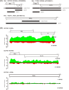
Fig 1. Genome sequencing of Groundnut chlorotic fan-spot virus (GCFSV) via RNA-seq.
(A) The contigs de novo assembled from transcriptomic reads (CqGCF) and viral RNA reads (GCF) were annotated to the reference sequences, for which the accession numbers are indicated. The codes of the contigs are indicated on the right side. The nucleotide positions of the individual open reading frames (ORFs, white boxes) in the reference RNAs are indicated. The ORFs encoded from the viral (v)-sense strand are shown on the upper side. The ORFs encoded from the viral complementary (vc)-sense strand are represented on the lower side. The contigs mapped to the references are shown as black boxes, and the non-mapped sequences are shown as gray boxes. (B) Analyses for the read coverage of reads mapping to the S, M and L RNAs of GCFSV. The green and red peaks represent the reads mapping to the v-sense strand and vc-sense strand of RNAs, respectively. The y-axis shows the reads coverage.
Fig 2
Fig 2. Phylogenetic analyses of the encoded proteins of the L and M RNAs of Groundnut chlorotic fan-spot virus (GCFSV).
(A) The conserved motifs of the RNA-dependent RNA polymerases (RdRps) of tospoviruses are compared, and the consensus sequences are shown in bold. The identical residues within the same motif are underlined. (B) Phylogenetic trees of RdRp, NSm and Gn/Gc precursor. The dendrograms were produced using the Neighbor-Joining algorithm with 1,000 bootstrap replicates. The percentages are shown. See Table 2for the virus names.
Fig 3
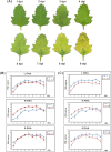
Fig 3. Progression of Groundnut chlorotic fan-spot virus (GCFSV) infection in Chenopodium quinoa leaves.
(A) The development of local lesions on the GCFSV-inoculated C. quinoa leaves was recorded from the first day post-inoculation (dpi) for 9 days at a 24-h interval. (B and C) The replication of GCFSV in the leaves of C. quinoa was analyzed by relative quantitation (RQ) assays. Total RNAs extracted from the leaves described in (A) were employed for quantitative real-time reverse transcription-polymerase chain reaction (qRT-PCR) amplification, using the primer pairs corresponding to the viral (v)-sense strand or viral complementary (vc)-sense strand of individual genomic RNAs of GCFSV. The transcripts of the NADH dehydrogenase subunit 5 (nad5) gene (B) and the glyceraldehyde 3-phosphate dehydrogenase (GAPDH) gene (C) of C. quinoa were amplified through qRT-PCR as the endogenous controls for the plants. The averaged CT values from individual amplifications were obtained from three independent runs, with a duplicate in each run. The RQ values were calculated from 2−ΔΔCT, ΔΔCT = (CTTarget–CTEndogenous)GCFSV-infected−(CTTarget–CTEndogenous)Mock-inoculated. The value of (CTTarget)Mock-inoculated was set as 35 for the calculations.
Fig 4
Fig 4. Analyses of the foliar transcriptome of Chenopodium quinoa.
(A) The workflow for the transcriptome analysis. Transcriptomic contigs de novo assembled from RNA-seq reads of mock-inoculated and Groundnut chlorotic fan-spot virus (GCFSV)-infected leaves of C. quinoa were annotated to obtain deduced complete genes. The fragments per kilobase of transcript per million mapped reads (FPKM) method was used to evaluate the differentiation of gene expression between the mock-inoculated and GCFSV-infected leaves of C. quinoa. The gene functions of the C. quinoa genes were deduced and classified using the Gene Ontology (GO), Kyoto Encyclopedia of Genes and Genomes (KEGG) and Interpro domain databases. (B) The pie chart shows the percentages of complete genes, partial genes and undefined contigs in the whole transcriptome. (C) The GO categories of biological processes (i), cellular components (ii) and molecular functions (iii) for the foliar transcriptome of GCFSV-infected C. quinoa at 4 days post-inoculation (dpi). The x-axis shows the number of contigs in each category. The y-axis indicates the name of a specific category of genes within that main category.
Fig 5
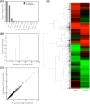
Fig 5. Statistical analysis of gene similarity between Chenopodium quinoa and Arabidopsis.
The length distribution (A) and nucleotide identity (B) of the coding sequences (CDS) of C. quinoa and Arabidopsis are shown. (C) Scatter plot of the CDS length in C. quinoa compared with that of Arabidopsis. (D) Relative expression patterns of mock-inoculated and Groundnut chlorotic fan-spot virus (GCFSV)-infected C. quinoa samples at 4 days post-inoculation (dpi). A relatively lower expression level is indicated in green color; a relatively higher expression level is indicated in red color; and no significant difference is indicated in black color. Genes with similar expression levels are grouped by lines.
Fig 6
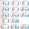
Fig 6. Relative quantitation (RQ) assays of gene modulation in the leaves of Chenopodium quinoa inoculated with Groundnut chlorotic fan-spot virus (GCFSV).
Hypersensitive response-related genes, including pathogenesis-related (PR) orthologues (A), transcription factor (TF) orthologues (B), mitogen-activated protein kinase (MAPK) orthologues (C), defense-related orthologues (D), and ethylene signaling-related orthologues (E) were assayed in C. quinoa using total RNAs extracted from the mock-inoculated and virus-infected leaf tissues at 1 day post-inoculation (dpi) and 4 dpi. The corresponding contig IDs of the assayed genes are indicated in parentheses. The glyceraldehyde 3-phosphate dehydrogenase (GAPDH) gene of C. quinoa was used as the comparative reference. The SYBR Green I-based one-step quantitative real-time reverse transcription-polymerase chain reaction (qRT-PCR) amplification and RQ calculation were performed using the 2−ΔΔCT method. Each RQ value was averaged from three independent runs, with a duplicate for each run. The means ± standard errors are presented. The RQ values of the mock-inoculated group were normalized to 1 for comparison in all assays. Significant differences between means compared with mock inoculation were determined using the Student’s t-test (*p < 0.05, **p < 0.01). The RQ results are shown in the blue columns (right side). The fragments per kilobase of transcript per million mapped reads (FPKM) results for each assayed gene are shown in the red columns (left side).

References

    1. Pappu HR, Jones RA, Jain RK. Global status of tospovirus epidemics in diverse cropping systems: Successes gained and challenges that lie ahead. Virus Res. 2009; 141:219–236. doi: 10.1016/j.virusres.2009.01.009 - DOI - PubMed
    1. Plyusnin A, Beaty BJ, Elliott RM, Goldbach R, Kormelink R, Lundkvist A, et al. Bunyaviridae In: King AMQ, Lefkowitz E, Adams MJ, Carstens EB, editors. Virus taxonomy: 9th report of the international committee on taxonomy of viruses. New York: Elsevier; 2011. pp. 725–740.
    1. de Haan P, Kormelink R, Resende RO, van Poelwijk F, Peters D, Goldbach R. Tomato spotted wilt virus L RNA encodes a putative RNA polymerase. J Gen Virol. 1991; 72:2207–2216. doi: 10.1099/0022-1317-72-9-2207 - DOI - PubMed
    1. van Knippenberg I, Goldbach R, Kormelink R. Purified Tomato spotted wilt virus particles support both genome replication and transcription in vitro. Virology. 2002; 303:278–286. - PubMed
    1. Kormelink R, Storms M, Van Lent J, Peters D, Goldbach R. Expression and subcellular location of the NSm protein of Tomato spotted wilt virus (TSWV), a putative viral movement protein. Virology. 1994; 200:56–65. doi: 10.1006/viro.1994.1162 - DOI - PubMed

LinkOut - more resources