

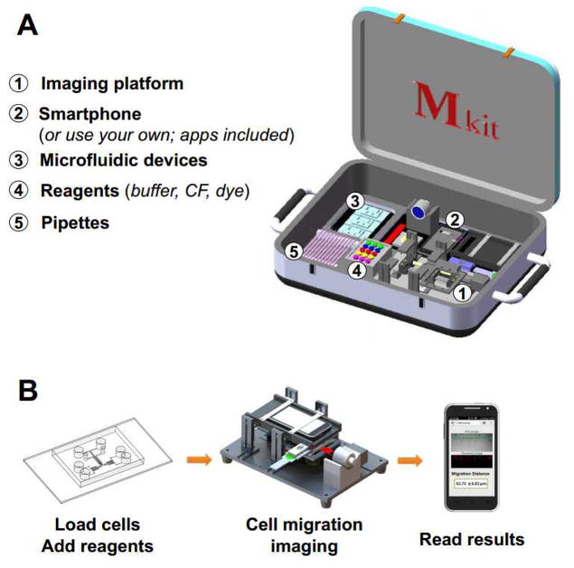
Mkit: A cell migration assay based on microfluidic device and smartphone
- PMID: 28772229
- PMCID: PMC5585005
- DOI: 10.1016/j.bios.2017.07.064
Mkit: A cell migration assay based on microfluidic device and smartphone
Abstract
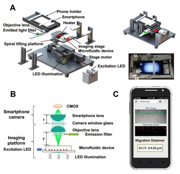
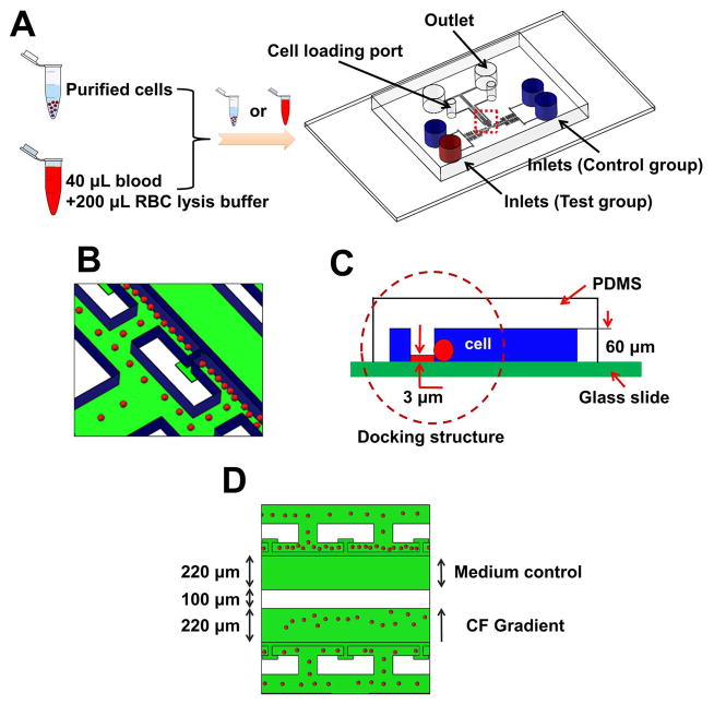
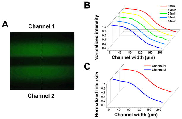
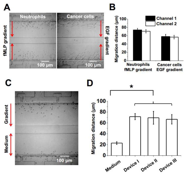
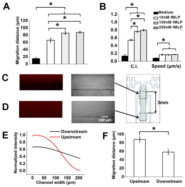
Mobile sensing based on the integration of microfluidic device and smartphone, so-called MS2 technology, has enabled many applications over recent years, and continues to stimulate growing interest in both research communities and industries. In particular, it has been envisioned that MS2 technology can be developed for various cell functional assays to enable basic research and clinical applications. Toward this direction, in this paper, we describe the development of a MS2-based cell functional assay for testing cell migration (the Mkit). The system is constructed as an integrated test kit, which includes microfluidic chips, a smartphone-based imaging platform, the phone apps for image capturing and data analysis, and a set of reagent and accessories for performing the cell migration assay. We demonstrated that the Mkit can effectively measure purified neutrophil and cancer cell chemotaxis. Furthermore, neutrophil chemotaxis can be tested from a drop of whole blood using the Mkit with red blood cell (RBC) lysis. The effects of chemoattractant dose and gradient profile on neutrophil chemotaxis were also tested using the Mkit. In addition to research applications, we demonstrated the effective use of the Mkit for on-site test at the hospital and for testing clinical samples from chronic obstructive pulmonary disease patient. Thus, this developed Mkit provides an easy and integrated experimental platform for cell migration related research and potential medical diagnostic applications.
Keywords: Cell functional assay; Cell migration; Chemotaxis; Microfluidic device; Smartphone.
Copyright © 2017 Elsevier B.V. All rights reserved.
Figures
References
-
- Albini A, Benelli R. Nat Protoc. 2007;2(3):504–511. - PubMed
-
- Arpa A, Wetzstein G, Lanman D, Raskar R. 2012 IEEE Computer Society Conference on Computer Vision and Pattern Recognition Workshops; IEEE; 2012. pp. 23–28.
-
- Chan HN, Shu Y, Xiong B, Chen Y, Chen Y, Tian Q, Michael SA, Shen B, Wu H. ACS Sensors. 2015;1(3):227–234.
MeSH terms
Grants and funding
LinkOut - more resources
Full Text Sources
Other Literature Sources
