

Improved Margins Detection of Regions Enriched with Gold Nanoparticles inside Biological Phantom
- PMID: 28772563
- PMCID: PMC5459194
- DOI: 10.3390/ma10020203
Improved Margins Detection of Regions Enriched with Gold Nanoparticles inside Biological Phantom
Abstract
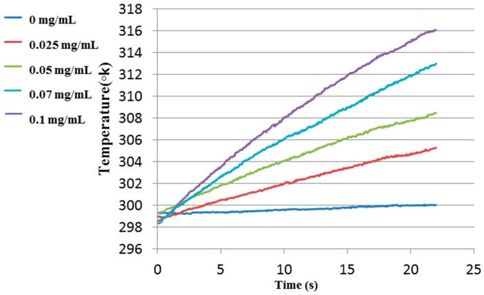
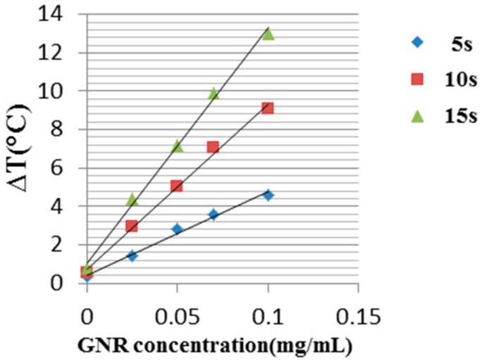
Utilizing the surface plasmon resonance (SPR) effect of gold nanoparticles (GNPs) enables their use as contrast agents in a variety of biomedical applications for diagnostics and treatment. These applications use both the very strong scattering and absorption properties of the GNPs due to their SPR effects. Most imaging methods use the light-scattering properties of the GNPs. However, the illumination source is in the same wavelength of the GNPs' scattering wavelength, leading to background noise caused by light scattering from the tissue. In this paper we present a method to improve border detection of regions enriched with GNPs aiming for the real-time application of complete tumor resection by utilizing the absorption of specially targeted GNPs using photothermal imaging. Phantoms containing different concentrations of GNPs were irradiated with a continuous-wave laser and measured with a thermal imaging camera which detected the temperature field of the irradiated phantoms. By modulating the laser illumination, and use of a simple post processing, the border location was identified at an accuracy of better than 0.5 mm even when the surrounding area got heated. This work is a continuation of our previous research.
Keywords: gold nanorods; laser beam modulation; photothermal imaging; surface plasmon resonance.
Conflict of interest statement
The authors declare no conflict of interest.
Figures
Similar articles
-
Towards real-time detection of tumor margins using photothermal imaging of immune-targeted gold nanoparticles.Int J Nanomedicine. 2012;7:4707-13. doi: 10.2147/IJN.S34157. Epub 2012 Aug 28. Int J Nanomedicine. 2012. PMID: 22956871 Free PMC article.
-
An ultra-sensitive dual-mode imaging system using metal-enhanced fluorescence in solid phantoms.Nano Res. 2015 Dec 1;8(12):3912-3921. doi: 10.1007/s12274-015-0891-y. Nano Res. 2015. PMID: 26870306 Free PMC article.
-
In vitro outlook of gold nanoparticles in photo-thermal therapy: a literature review.Lasers Med Sci. 2018 May;33(4):917-926. doi: 10.1007/s10103-018-2467-z. Epub 2018 Feb 28. Lasers Med Sci. 2018. PMID: 29492712 Review.
-
Low power argon laser-induced thermal therapy for subcutaneous Ehrlich carcinoma in mice using spherical gold nanoparticles.J Biomed Nanotechnol. 2010 Dec;6(6):687-93. doi: 10.1166/jbn.2010.1166. J Biomed Nanotechnol. 2010. PMID: 21361134
-
Laser-induced optothermal response of gold nanoparticles: From a physical viewpoint to cancer treatment application.J Biophotonics. 2021 Feb;14(2):e202000161. doi: 10.1002/jbio.202000161. Epub 2020 Nov 3. J Biophotonics. 2021. PMID: 32761778 Review.
Cited by
-
An Engineered Nanocomplex with Photodynamic and Photothermal Synergistic Properties for Cancer Treatment.Int J Mol Sci. 2022 Feb 18;23(4):2286. doi: 10.3390/ijms23042286. Int J Mol Sci. 2022. PMID: 35216400 Free PMC article.
-
Gold Nanorods for Doxorubicin Delivery: Numerical Analysis of Electric Field Enhancement, Optical Properties and Drug Loading/Releasing Efficiency.Materials (Basel). 2022 Feb 26;15(5):1764. doi: 10.3390/ma15051764. Materials (Basel). 2022. PMID: 35268995 Free PMC article.
-
Innovative functional polymerization of pyrrole-N-propionic acid onto WS2 nanotubes using cerium-doped maghemite nanoparticles for photothermal therapy.Sci Rep. 2021 Sep 23;11(1):18883. doi: 10.1038/s41598-021-97052-6. Sci Rep. 2021. PMID: 34556680 Free PMC article.
-
Small Gold Nanorods: Recent Advances in Synthesis, Biological Imaging, and Cancer Therapy.Materials (Basel). 2017 Nov 30;10(12):1372. doi: 10.3390/ma10121372. Materials (Basel). 2017. PMID: 29189739 Free PMC article. Review.
References
-
- Kircher M.F., de la Zerda A., Jokerst J.V., Zavaleta C.L., Kempen P.J., Mittra E., Pitter K., Huang R., Campos C., Habte F., et al. A brain tumor molecular imaging strategy using a new triple-modality MRI-photoacoustic-Raman nanoparticle. Nat. Med. 2012;18:829–834. doi: 10.1038/nm.2721. - DOI - PMC - PubMed
-
- Van Dam G.M., Themelis G., Crane L.M., Harlaar N.J., Pleijhuis R.G., Kelder W., Sarantopoulos A., de Jong J.S., Arts H.J., van der Zee A.G., et al. Intraoperative tumor-specific fluorescence imaging in ovarian cancer by folate receptor-α targeting: First in-human results. Nat. Med. 2011;17:1315–1319. doi: 10.1038/nm.2472. - DOI - PubMed
LinkOut - more resources
Full Text Sources
Other Literature Sources
