. 2017 Mar 14;10(3):288.
doi: 10.3390/ma10030288.

Cytotoxicity of Light-Cured Dental Materials according to Different Sample Preparation Methods

Affiliations

Cytotoxicity of Light-Cured Dental Materials according to Different Sample Preparation Methods

Myung-Jin Lee et al. Materials (Basel). .

Abstract

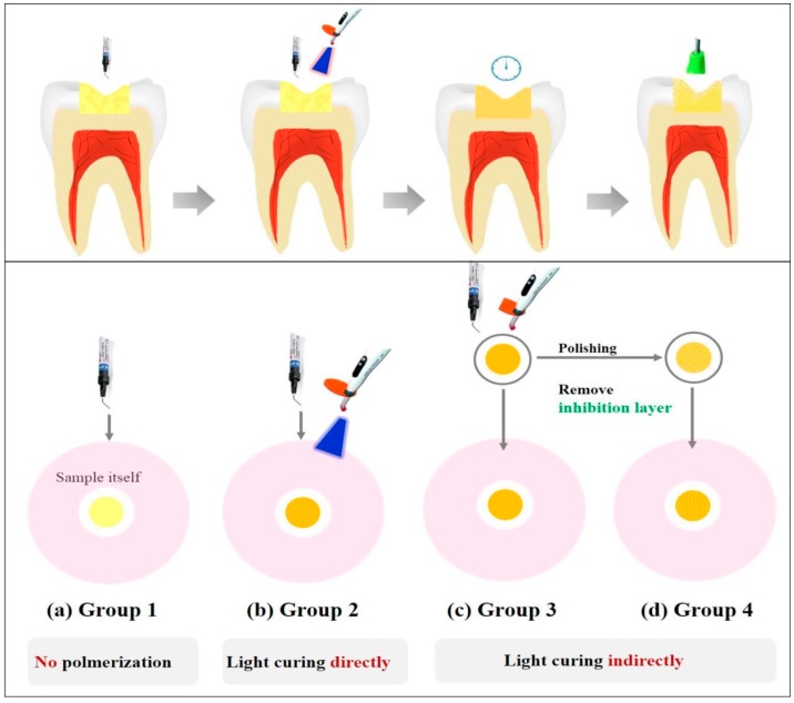
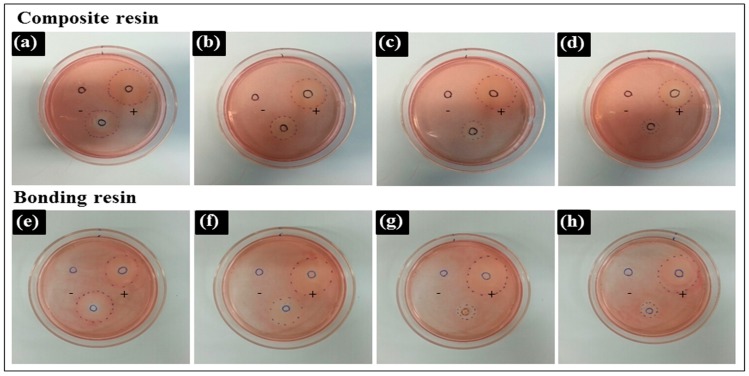
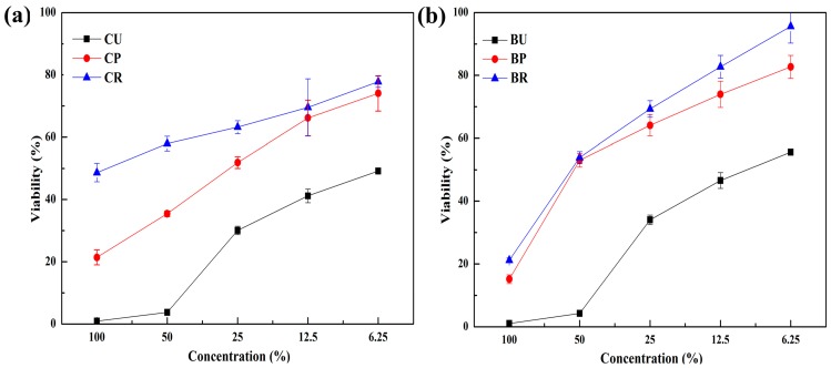
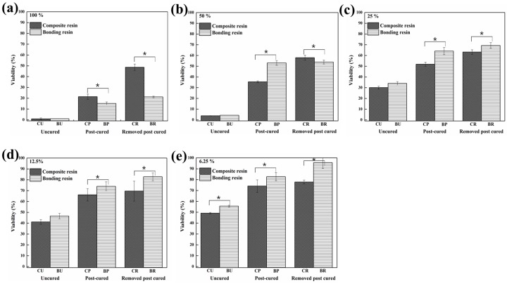
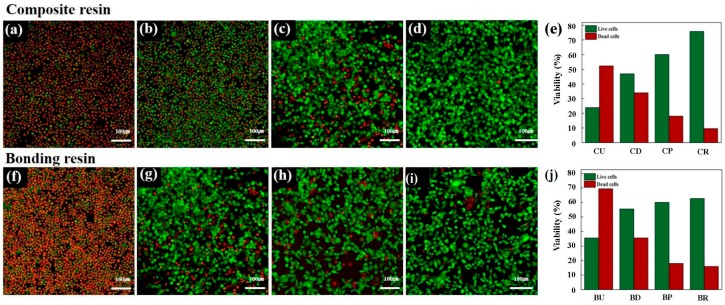
Dental light-cured resins can undergo different degrees of polymerization when applied in vivo. When polymerization is incomplete, toxic monomers may be released into the oral cavity. The present study assessed the cytotoxicity of different materials, using sample preparation methods that mirror clinical conditions. Composite and bonding resins were used and divided into four groups according to sample preparation method: uncured; directly cured samples, which were cured after being placed on solidified agar; post-cured samples were polymerized before being placed on agar; and "removed unreacted layer" samples had their oxygen-inhibition layer removed after polymerization. Cytotoxicity was evaluated using an agar diffusion test, MTT assay, and confocal microscopy. Uncured samples were the most cytotoxic, while removed unreacted layer samples were the least cytotoxic (p < 0.05). In the MTT assay, cell viability increased significantly in every group as the concentration of the extracts decreased (p < 0.05). Extracts from post-cured and removed unreacted layer samples of bonding resin were less toxic than post-cured and removed unreacted layer samples of composite resin. Removal of the oxygen-inhibition layer resulted in the lowest cytotoxicity. Clinicians should remove unreacted monomers on the resin surface immediately after restoring teeth with light-curing resin to improve the restoration biocompatibility.

Keywords: biocompatibility; cytotoxicity; oxygen-inhibition layer; resin-based materials; sample preparation.


Conflict of interest statement

The authors declare no conflict of interest.

Figures

Figure 1
Figure 1
A representative illustration of the different sample preparation methods. (a) Samples were used without any curing; (b) samples were cured on agar with a light curing unit for 20 s; (c) samples were used after polymerization; (d) samples were polymerized and the unreacted resin monomer layer was removed afterward.
Figure 2
Figure 2
Decolorization zones of experimental materials in the agar diffusion test. The empty Teflon mold (negative control), the latex sheet from the latex glove (positive control), and experimental samples of (a) uncured composite resin (CU); (b) directly cured composite resin (CD); (c) post-cured composite resin (CP); (d) composite resin after removing the unreacted layer (CR); (e) uncured bonding resin (BU); (f) directly cured bonding resin (BD); (g) post-cured bonding resin (BP); (h) bonding resin after removing the unreacted layer (BR) were located in predetermined positions. Representative images are shown after experiments were performed in triplicate.
Figure 3
Figure 3
Cell viability following exposure to the extracts from (a) composite resin and (b) bonding resin at different dilutions. B: bonding resin; C: composite resin; U: uncured; P: post-cured; R: unreacted layer removed.
Figure 4
Figure 4
Cell viability following exposure to the extracts from the composite resin and bonding resin at different dilutions: (a) 100% concentration; (b) 50% concentration; (c) 25% concentration; (d) 12.5% concentration; (e) 6.25% concentration (*: Statistically significant at p < 0.05).
Figure 5
Figure 5
Confocal laser microscopy images following calcein AM and ethidium homodimer-1 staining of L929 cells. Cells were exposed to extracts from (a) uncured composite resin; (b) directly cured composite resin; (c) post-cured composite resin; (d) composite resin after removing the unreacted layer; (e) Live/Dead Assay® quantified the live and dead cells in equivalent surface areas of composite resin; (f) uncured bonding resin; (g) directly cured bonding resin; (h) post-cured bonding resin; (i) bonding resin after removing the unreacted layer; (j) Live/Dead Assay® quantified the live and dead cells in equivalent surface areas of bonding resin. Live cells are stained green and dead cells are stained red for confocal laser microscope images.

Similar articles

Cited by

References

    1. Xia Y., Zhang F., Xie H., Gu N. Nanoparticle-reinforced resin-based dental composites. J. Dent. 2008;36:450–455. doi: 10.1016/j.jdent.2008.03.001. - DOI - PubMed
    1. Jandt K.D., Sigusch B.W. Future perspectives of resin-based dental materials. Dent. Mater. 2009;25:1001–1006. doi: 10.1016/j.dental.2009.02.009. - DOI - PubMed
    1. Chai Z., Li F., Fang M., Wang Y., Ma S., Xiao Y., Huang L., Chen J. The bonding property and cytotoxicity of a dental adhesive incorporating a new antibacterial monomer. J. Oral Rehabil. 2011;38:849–856. doi: 10.1111/j.1365-2842.2011.02212.x. - DOI - PubMed
    1. Sigusch B.W., Pflaum T., Volpel A., Schinkel M., Jandt K.D. The influence of various light curing units on the cytotoxicity of dental adhesives. Dent. Mater. 2009;25:1446–1452. doi: 10.1016/j.dental.2009.06.016. - DOI - PubMed
    1. Sigusch B.W., Volpel A., Braun I., Uhl A., Jandt K.D. Influence of different light curing units on the cytotoxicity of various dental composites. Dent. Mater. 2007;23:1342–1348. doi: 10.1016/j.dental.2006.11.013. - DOI - PubMed

LinkOut - more resources