Review
. 2017 Mar 24;10(4):334.
doi: 10.3390/ma10040334.

Calcium Phosphate Bioceramics: A Review of Their History, Structure, Properties, Coating Technologies and Biomedical Applications

Affiliations
Review

Calcium Phosphate Bioceramics: A Review of Their History, Structure, Properties, Coating Technologies and Biomedical Applications

Noam Eliaz et al. Materials (Basel). .

Abstract

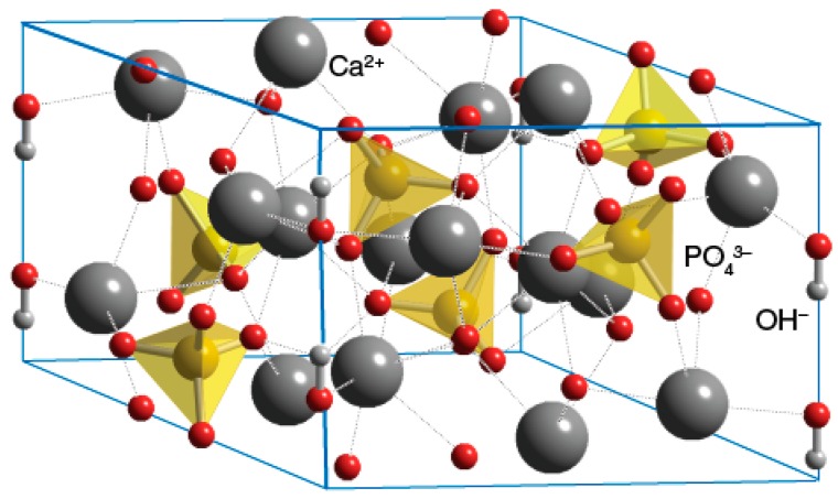
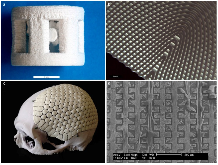
Calcium phosphate (CaP) bioceramics are widely used in the field of bone regeneration, both in orthopedics and in dentistry, due to their good biocompatibility, osseointegration and osteoconduction. The aim of this article is to review the history, structure, properties and clinical applications of these materials, whether they are in the form of bone cements, paste, scaffolds, or coatings. Major analytical techniques for characterization of CaPs, in vitro and in vivo tests, and the requirements of the US Food and Drug Administration (FDA) and international standards from CaP coatings on orthopedic and dental endosseous implants, are also summarized, along with the possible effect of sterilization on these materials. CaP coating technologies are summarized, with a focus on electrochemical processes. Theories on the formation of transient precursor phases in biomineralization, the dissolution and reprecipitation as bone of CaPs are discussed. A wide variety of CaPs are presented, from the individual phases to nano-CaP, biphasic and triphasic CaP formulations, composite CaP coatings and cements, functionally graded materials (FGMs), and antibacterial CaPs. We conclude by foreseeing the future of CaPs.

Keywords: bioceramics; biomineralization; bone cement; calcium phosphate; coating; composites; drug delivery; electrochemical deposition; functionally graded materials; nano-hydroxyapatite.


Conflict of interest statement

The authors declare no conflict of interest.

Figures

Figure 1
Figure 1
(a) Hierarchical structure of bone [88]. Reproduced with permission from Nature Publishing Group; (b) Typical structure of long bone [91]. Reproduced from Tortora and Derrickson, Principles of Anatomy and Physiology, 11th edition, © John Wiley & Sons, Inc.
Figure 2
Figure 2
(a) Ashby chart of strength versus Young’s modules of elasticity (specific values) for natural and synthetic materials. Note values for collagen, hydroxyapatite (HAp), cancellous bone, compact bone and enamel; (b) Projections for natural and synthetic materials [88]. Reproduced with permission from Nature Publishing Group.
Figure 3
Figure 3
Thermodynamic calculations, using the PHREEQC software, that predict which of five different CaP phases may precipitate spontaneously in electrolyte solutions at different ion concentrations and pH values. In all cases the bath temperature is 37 °C: (a) X0.1 bath; (b) X10 bath; and (c) Nominal bath [191]. Reproduced with permission from Elsevier B.V.
Figure 4
Figure 4
Equilibrium phase diagram of different calcium phosphates. The shaded region shows the phases of interest for biphasic calcium phosphate (BCP) formation (T1 = 1360 °C, T2 = 1475 °C) [245]. In this figure, TTCP—tetracalcium phosphate, CaO—calcium oxide. Reproduced with permission of Elsevier Ltd.
Figure 5
Figure 5
Unit cell of hexagonal HAp (space group P63/m) [375]. Reproduced with permission from ChemTube3D, The University of Liverpool.
Figure 6
Figure 6
Schematic illustration of the novel approach for electrochemical deposition of pure HAp NPs for coating dental implants suggested by Mandler, Eliaz et al. in Reference s [424,425].
Figure 7
Figure 7
SEM images of MBA-15 osteogenic cells on surfaces of: (a) Gr-Ti; (b,f) HAp4.2; and (ce,g) HAp6.0 [238]. Reproduced with permission from Elsevier Ltd.
Figure 8
Figure 8
A widened hole in the medullary canal of the distal femur of a rabbit before (a) and after (b) press fitting of the implant; (c,d) Radiographs of the right distal femur of a rabbit (c—Anterior-Posterior, AP, d—Lateral, LAT). The implant is press fitted into the medullar canal, within both the metaphysis and diaphysis [92]. Reproduced with permission from Elsevier Ltd.
Figure 9
Figure 9
(a) Average bone apposition ratio (BAR) after 7 and 14 days of implantation. The error bars are standard deviations (n = 3); (b) Aqueous solubilities of plasma-sprayed hydroxyapatite (PSHA) and electrochemically deposited hydroxyapatite (EDHA) in deionized water at room temperature. Bare Ti–6Al–4V alloy serves as a reference (n = 1; error bars are standard deviations from three measurements) [189]. Reproduced with permission from Elsevier Ltd.
Figure 10
Figure 10
The distribution of phosphate species as a function of pH at 37 °C, 0.36 mM total analytical concentration of phosphate, free hydrogen concentration of 10−6 M. The values of the three dissociation constants are marked, along with the CaP phases that are likely to form from each species. As the pH increases, the Ca/P ratio in the solid phase increases, and the solubility of this phase decreases [49], both in vitro and in vivo.
Figure 11
Figure 11
(a) Schematic diagram for the synthesis of CaP coating on flexible carbon-based membranes by electrodeposition; (b) Current transients of three types of membranes during the deposition process [805]. Reproduced with permission from Wiley-VCH Verlag GmbH and Co. KGaA, Weinheim.
Figure 12
Figure 12
The tensile stress to failure of ED HAp-coated samples with different pre-treatments during adhesion test. The data is presented in terms of mean + standard error of the mean (n = 3). The red dash line defines the minimum adhesion strength required by the US FDA for coating adhesion strength in orthopedic and dental endosseous implants [237]. Reproduced with permission from Springer Science+Business Media, LLC.
Figure 13
Figure 13
(ac) SEM images revealing the typical surface morphologies of electrochemically deposited hydroxyapatite (ED Hap) on ground Ti, Ti soaked in NaOH, and Ti soaked in H2O2, respectively; (d) High-magnification image of (b), which reveals the hexagonal cross-section of the bars; (e) The typical current density transients during potentiostatic deposition of HAp on the three types of substrate; (f) Cell density on different surfaces (partial population). The data are presented as mean ± standard deviation. Inset: Two typical fluorescent images of cell nuclei (Hoechst staining) on Gr-Ti vs. NaOH-Ti-HAp [238]. Reproduced with permission from Elsevier Ltd.
Figure 13
Figure 13
(ac) SEM images revealing the typical surface morphologies of electrochemically deposited hydroxyapatite (ED Hap) on ground Ti, Ti soaked in NaOH, and Ti soaked in H2O2, respectively; (d) High-magnification image of (b), which reveals the hexagonal cross-section of the bars; (e) The typical current density transients during potentiostatic deposition of HAp on the three types of substrate; (f) Cell density on different surfaces (partial population). The data are presented as mean ± standard deviation. Inset: Two typical fluorescent images of cell nuclei (Hoechst staining) on Gr-Ti vs. NaOH-Ti-HAp [238]. Reproduced with permission from Elsevier Ltd.
Figure 14
Figure 14
Various applications and forms of commercially available CaP-related products. (a) Bone augmentation after extraction of the left central incisro tooth. Courtesy Dr. Eyal Tarazi, DMD, Caesarea, Israel; (b) Coated dental implant. Reproduced with permission from SGS Dental Implant System [842]; (c) Augmentos® 3D Scaffold bone substitute material [843] for filling or reconstructing non-load-bearing bone defects or for filling bone defects that are sufficiently stabilized by appropriate means. This seems to be the first 3D-printed CaP cement. The extrusion printing process does not involve any heat treatment steps. Reproduced with permission from InnoTERE GmbH; (d) Calcibon® self-setting cement granules consisted of α-TCP, CaHPO4, CaCO3 and HAp [844]; (e) Megasonex® Nano-Hydroxyapatite Toothpaste [845]. This is the world’s first nano-HAp toothpaste designed specifically for electric and ultrasonic toothbrushes. Nano-HAp helps to safely remineralize enamel (potentially reversing early stage tooth decay, white spot caries) and encrusts harmful bacteria (helping to prevent plaque formation). Other ingredients include tetrasodium pyrophosphate (prevents plaque from sticking), sorbitol, xylitol, mica, titanium oxide, citric acid, sodium carboxymethylcellulose, sodium saccharin, glycerin and silica. This toothpaste is free of fluoride and undesirable foaming agents such as sodium lauryl sulfate (SLS). Reproduced with permission from Goldspire Group, Ltd. The first toothpaste containing synthetic HAp as an alternative to fluoride for the remineralization and reparation of tooth enamel, BioRepair® [846], appeared in Europe in 2006. The biomimetic zinc HAp (named microRepair®) is intended to protect the teeth by creating a new layer of synthetic enamel around the tooth instead of hardening the existing layer with fluoride that chemically changes it into fluorapatite; (f) Osteovit® xenograft bone substitute [847]. Reproduced with permission © B. Braun Melsungen AG; (g) DePuy Synthes CORAIL® cementless hip prosthesis for total hip arthroplasty [848]; (h) DePuy Synthes DBX™ Material bone graft substitute composed of demineralized bone matrix (DBM) from human donors in a sodium hyaluronate carrier [849].
Figure 15
Figure 15
Versatility, scalability, and manipulation of 3D-printed hyperelastic “bone” (HB). (A) Easy to synthesize volumes (~100 mL shown) of liquid-based HB inks (inset) can be 3D-printed into a variety of structures: 3D-printed 12 × 12 cm HAp-poly(d,l-lactic-co-glycolide acid) (PLGA) sheet comprising three layers, which can be manipulated in a variety of ways, including rolling, folding, and cutting. Origami methods may be used to create complex folded structures, whereas Kirigami methods can produce complex structures from strategic folding and cutting; (B) Full-scale, anatomically correct parts, such as a human mandible, comprising >250 layers, can be designed, 3D-printed from HAp-PLGA, and washed to rapidly produce a ready-to-implant object. Final image shows 3D-printed mandible next to an adult cadaveric human mandible; (C) Photograph series illustrating that custom-sized HAp-PLGA sleeves can be snuggly stretched around, cut, and sutured to a soft tissue, such as human cadaveric tendon, facilitating arthroscopic ACL repair and replacement surgery; (D) Independently 3D-printed HAp-PLGA miniature-scale versions of a human skull, skull cap, mandible, and upper thoracic seamlessly fused together to create highly complex structures by using HB ink applied to points of contact; (E) Black light-illuminated optical photographs of the outside and internal cross-sections of HAp-PLGA fibre with (top) and without (bottom) incorporated recombinant green fluorescent protein (rGFP) [862]. Reproduced with permission from The American Association for the Advancement of Science.
Figure 16
Figure 16
Examples of structures obtained by additive manufacturing techniques. (a) 3D printed scaffolds made of dicalcium phosphate anhydrous (DCPA) (scale bar: 5 mm) [864]; (b) Solid obtained by robocasting (scale bar: 2 mm); (c) “Craniomosaic”: a DCPA-based implant for treatment of cranial bone defects. The device uses a 3D-printed titanium mesh covered with DCPA ceramic tiles; (d) Pattern of CaP created on a silicon substrate using soft lithography (scale bar: 200 μm) [863]. Reproduced with permission from Elsevier Ltd.

Similar articles

Cited by

References

    1. Dorozhkin S.V. A detailed history of calcium orthophosphates from 1770s till 1950. Mater. Sci. Eng. C. 2013;33:3085–3110. doi: 10.1016/j.msec.2013.04.002. - DOI - PubMed
    1. Driskell T.D. Early history of calcium phosphate materials and coatings. In: Horowitz E., Parr J.E., editors. Characterization and Performance of Calcium Phosphate Coatings for Implants. American Society for Testing and Materials (ASTM); Philadelphia, PA, USA: 1994. pp. 1–9.
    1. Shackelford J.F. Bioceramics—An historical perspective. Mater. Sci. Forum. 1999;293:1–4. doi: 10.4028/www.scientific.net/MSF.293.1. - DOI
    1. Shepperd J. The early biological history of calcium phosphates. In: Epinette J.A., Manley M.T., editors. Fifteen Years of Clinical Experience with Hydroxyapatite Coatings in Joint Arthroplasty. Springer; Paris, France: 2004. pp. 3–8.
    1. Leeuwenhoek A. Microscopical observations concerning blood, milk, bone, the brain, spittle, and cuticula, etc. Philos. Trans. 1674;9:121–128. doi: 10.1098/rstl.1674.0030. - DOI