

Fabrication of Biocompatible Potassium Sodium Niobate Piezoelectric Ceramic as an Electroactive Implant
- PMID: 28772704
- PMCID: PMC5506920
- DOI: 10.3390/ma10040345
Fabrication of Biocompatible Potassium Sodium Niobate Piezoelectric Ceramic as an Electroactive Implant
Abstract
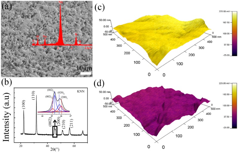
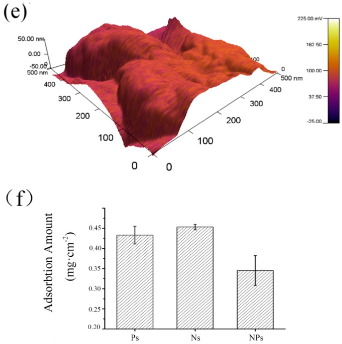
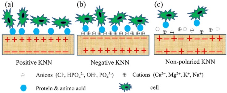
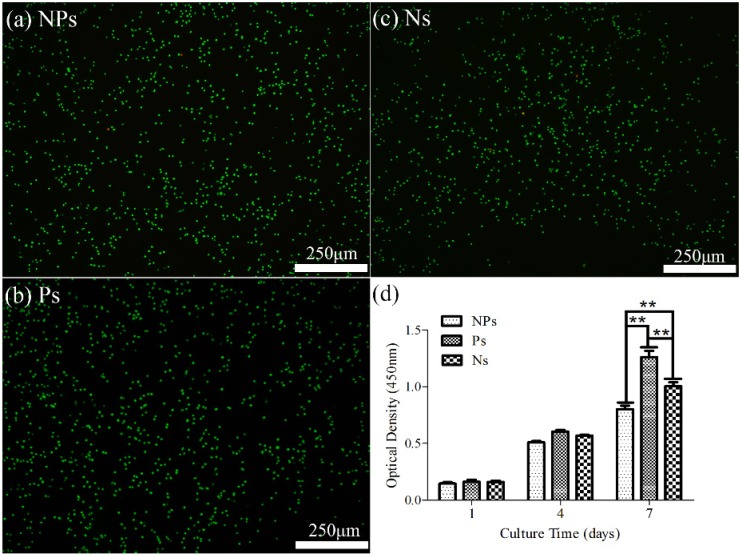
The discovery of piezoelectricity in natural bone has attracted extensive research in emulating biological electricity for various tissue regeneration. Here, we carried out experiments to build biocompatible potassium sodium niobate (KNN) ceramics. Then, influence substrate surface charges on bovine serum albumin (BSA) protein adsorption and cell proliferation on KNN ceramics surfaces was investigated. KNN ceramics with piezoelectric constant of ~93 pC/N and relative density of ~93% were fabricated. The adsorption of protein on the positive surfaces (Ps) and negative surfaces (Ns) of KNN ceramics with piezoelectric constant of ~93 pC/N showed greater protein adsorption capacity than that on non-polarized surfaces (NPs). Biocompatibility of KNN ceramics was verified through cell culturing and live/dead cell staining of MC3T3. The cells experiment showed enhanced cell growth on the positive surfaces (Ps) and negative surfaces (Ns) compared to non-polarized surfaces (NPs). These results revealed that KNN ceramics had great potential to be used to understand the effect of surface potential on cells processes and would benefit future research in designing piezoelectric materials for tissue regeneration.
Keywords: biological; electroactive; implant; piezoelectric; potassium sodium niobate.
Conflict of interest statement
The authors declare no conflict of interest.
Figures
Similar articles
-
The antibacterial effect of potassium-sodium niobate ceramics based on controlling piezoelectric properties.Colloids Surf B Biointerfaces. 2019 Mar 1;175:463-468. doi: 10.1016/j.colsurfb.2018.12.022. Epub 2018 Dec 12. Colloids Surf B Biointerfaces. 2019. PMID: 30572154
-
High-Performance 0-3 Type Niobate-Based Lead-Free Piezoelectric Composite Ceramics with ZnO Inclusions.ACS Appl Mater Interfaces. 2018 Sep 12;10(36):30566-30573. doi: 10.1021/acsami.8b10136. Epub 2018 Aug 27. ACS Appl Mater Interfaces. 2018. PMID: 30107108
-
Hardening Effect in Lead-Free KNN-Based Piezoelectric Ceramics with CuO Doping.ACS Appl Mater Interfaces. 2022 Dec 21;14(50):55803-55811. doi: 10.1021/acsami.2c18015. Epub 2022 Dec 8. ACS Appl Mater Interfaces. 2022. PMID: 36482677
-
Sintering of Lead-Free Piezoelectric Sodium Potassium Niobate Ceramics.Materials (Basel). 2015 Dec 1;8(12):8117-8146. doi: 10.3390/ma8125449. Materials (Basel). 2015. PMID: 28793702 Free PMC article. Review.
-
Exploring the application of piezoelectric ceramics in bone regeneration.J Biomater Appl. 2024 Nov;39(5):409-420. doi: 10.1177/08853282241274528. Epub 2024 Aug 17. J Biomater Appl. 2024. PMID: 39152927 Review.
Cited by
-
Processing Optimization and Toxicological Evaluation of "Lead-Free" Piezoceramics: A KNN-Based Case Study.Materials (Basel). 2021 Aug 3;14(15):4337. doi: 10.3390/ma14154337. Materials (Basel). 2021. PMID: 34361531 Free PMC article.
-
The Osteogenic Role of Barium Titanate/Polylactic Acid Piezoelectric Composite Membranes as Guiding Membranes for Bone Tissue Regeneration.Int J Nanomedicine. 2022 Sep 17;17:4339-4353. doi: 10.2147/IJN.S378422. eCollection 2022. Int J Nanomedicine. 2022. PMID: 36160471 Free PMC article.
-
Piezoelectricity Regulating Immune Osteogenesis in Osteoporosis.BME Front. 2025 Jul 2;6:0146. doi: 10.34133/bmef.0146. eCollection 2025. BME Front. 2025. PMID: 40606524 Free PMC article.
-
3D printing of piezoelectric and bioactive barium titanate-bioactive glass scaffolds for bone tissue engineering.Mater Today Bio. 2023 Jul 6;21:100719. doi: 10.1016/j.mtbio.2023.100719. eCollection 2023 Aug. Mater Today Bio. 2023. PMID: 37529217 Free PMC article.
-
Piezoelectric Electrospun Fibrous Scaffolds for Bone, Articular Cartilage and Osteochondral Tissue Engineering.Int J Mol Sci. 2022 Mar 8;23(6):2907. doi: 10.3390/ijms23062907. Int J Mol Sci. 2022. PMID: 35328328 Free PMC article. Review.
References
-
- Fukada E., Yasuda I. On the piezoelectric effect of bone. J. Phys. Soc. Jpn. 1957;12:1158–1162. doi: 10.1143/JPSJ.12.1158. - DOI
-
- Eiichi F., Iwao Y. Piezoelectric effects in collagen. Jpn. J. Appl. Phys. 1964;3:117.
LinkOut - more resources
Full Text Sources
Other Literature Sources
Miscellaneous
