Review
. 2017 May 6;10(5):507.
doi: 10.3390/ma10050507.

Current Insights into the Modulation of Oral Bacterial Degradation of Dental Polymeric Restorative Materials

Affiliations
Review

Current Insights into the Modulation of Oral Bacterial Degradation of Dental Polymeric Restorative Materials

Ning Zhang et al. Materials (Basel). .

Abstract

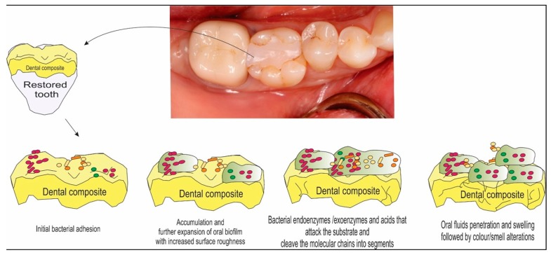
Dental polymeric composites have become the first choice for cavity restorations due to their esthetics and capacity to be bonded to the tooth. However, the oral cavity is considered to be harsh environment for a polymeric material. Oral biofilms can degrade the polymeric components, thus compromising the marginal integrity and leading to the recurrence of caries. Recurrent caries around restorations has been reported as the main reason for restoration failure. The degradation of materials greatly compromises the clinical longevity. This review focuses on the degradation process of resin composites by oral biofilms, the mechanisms of degradation and its consequences. In addition, potential future developments in the area of resin-based dental biomaterials with an emphasis on anti-biofilm strategies are also reviewed.

Keywords: biofilm; degradation; dental caries; dental materials.


Conflict of interest statement

The authors declare no conflict of interest.

Figures

Figure 1
Figure 1
Schematic illustration of a composite surface representing the clinical view of a tooth restored with dental restorative esthetic materials. This sequence of drawings shows the complex interactions between the material’s surface and the biofilm formation over time.
Figure 2
Figure 2
Scanning electron microscope (SEM) image of resin composite covered by oral biofilm (5000×). Observe the abundant bacterial colonies with many streptococcal chains (narrows). S. mutans, which composes a significant proportion of the oral streptococci in caries lesions, has been identified as the major etiological agent of human dental caries.
Figure 3
Figure 3
Representative transmission electron microscopy (TEM) micrographs representing the size and dispersion of silver nanoparticles (NAg) in a resin matrix: (A) lower and (B) higher magnifications. The NAg were formed in the resin by simultaneous reduction of the silver salt and photopolymerization of the dimethacrylates. Arrows indicate the silver nanoparticles, which were well dispersed in the resin with minimal appearance of nanoparticle aggregates. Adapted with permission from [36], copyright SAGE publications, 2012.
Figure 4
Figure 4
SEM micrographs of plaque microcosm biofilms: (A,B) lower magnification and (C,D) higher magnification. In (A), the composite control was covered with dense biofilms consisting of numerous long strings (arrows). In (B), Quaternary ammonium dimetacrylate associated to nanoparticles of amorphous calcium phosphate (NACP-QADM) nanocomposites had thinner biofilms with numerous pores “P”, without long strings. In (C), the long strings were made of bacterial cells connected with each other, and the cells had a normal, healthy short-rod morphology. However, as shown in (D), many cells on NACP-QADM nanocomposites had dissolved into pieces, while other cells still had a normal short-rod shape (long arrows indicate cell disintegration, and short arrows indicate normal healthy cells). Adapted with permission from [29], copyright Elsevier 2013.
Figure 5
Figure 5
Representative live/dead staining images of biofilms adherent on composite disks cultured for two days: (A) commercial control composite; (B) control composite with 0% 2-methacryloyloxyethyl phosphorylcholine (MPC); (C) control composite with 1.5% MPC and (D) control composite with 3% MPC. The live bacteria were stained green, and the dead bacteria were stained red. When live and dead bacteria were in close proximity or on the top of each other, the staining had yellow or orange colors. The composite disks had primarily live bacteria, with few dead bacteria. The commercial control composite (A) and the control composite with 0% MPC (B) had noticeably more bacteria coverage than composites containing MPC. There was less biofilm coverage on control composite disks containing MPC (C,D). The control composite with 3% MPC (D) had the least biofilm coverage. Adapted with permission from [52], copyright Nature Publishing Group 2013.

References

    1. Ferracane J.L. Resin composite—State of the art. Dent. Mater. 2011;27:29–38. doi: 10.1016/j.dental.2010.10.020. - DOI - PubMed
    1. Drummond J.L. Degradation, fatigue, and failure of resin dental composite materials. J. Dent. Res. 2008;87:710–719. doi: 10.1177/154405910808700802. - DOI - PMC - PubMed
    1. Bayne S.C. Correlation of clinical performance with ‘in vitro tests’ of restorative dental materials that use polymer-based matrices. Dent. Mater. 2012;28:52–71. doi: 10.1016/j.dental.2011.08.594. - DOI - PubMed
    1. Beyth N., Bahir R., Matalon S., Domb A.J., Weiss E.I. Streptococcus mutans biofilm changes surface-topography of resin composites. Dent. Mater. 2008;24:732–736. - PubMed
    1. Beyth N., Domb A.J., Weiss E.I. An in vitro quantitative antibacterial analysis of amalgam and composite resins. J. Dent. 2007;35:201–206. doi: 10.1016/j.jdent.2006.07.009. - DOI - PubMed

LinkOut - more resources