. 2017 May 31;10(6):606.
doi: 10.3390/ma10060606.

Graphene Nanosheets to Improve Physico-Mechanical Properties of Bioactive Calcium Silicate Cements

Affiliations

Graphene Nanosheets to Improve Physico-Mechanical Properties of Bioactive Calcium Silicate Cements

Nileshkumar Dubey et al. Materials (Basel). .

Abstract

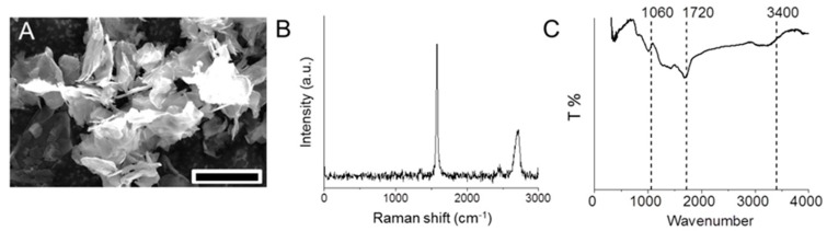
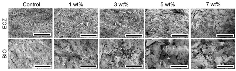
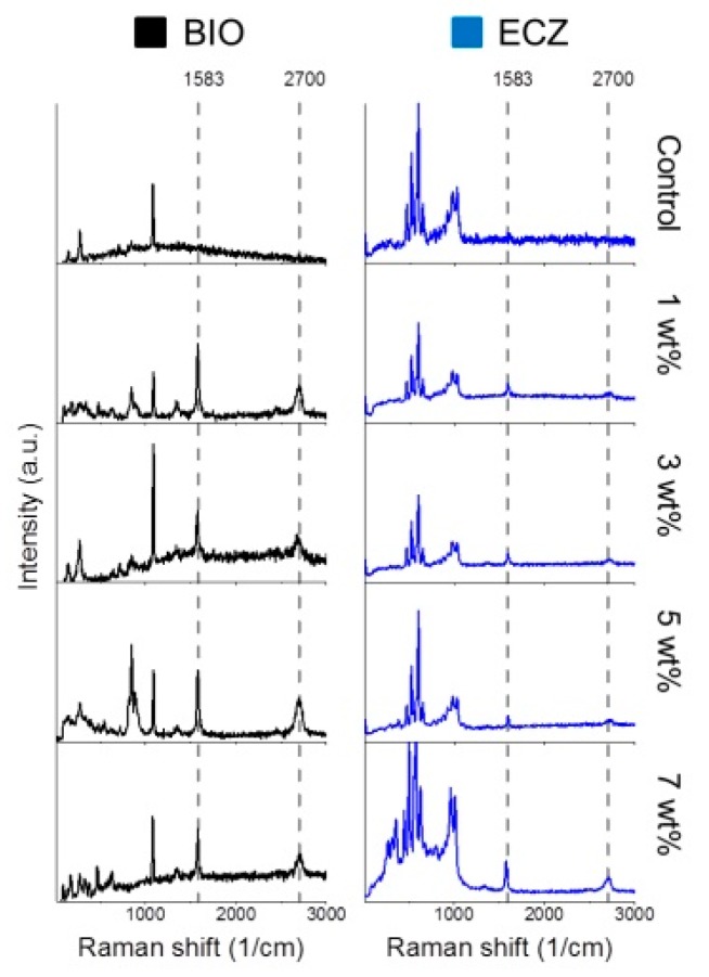
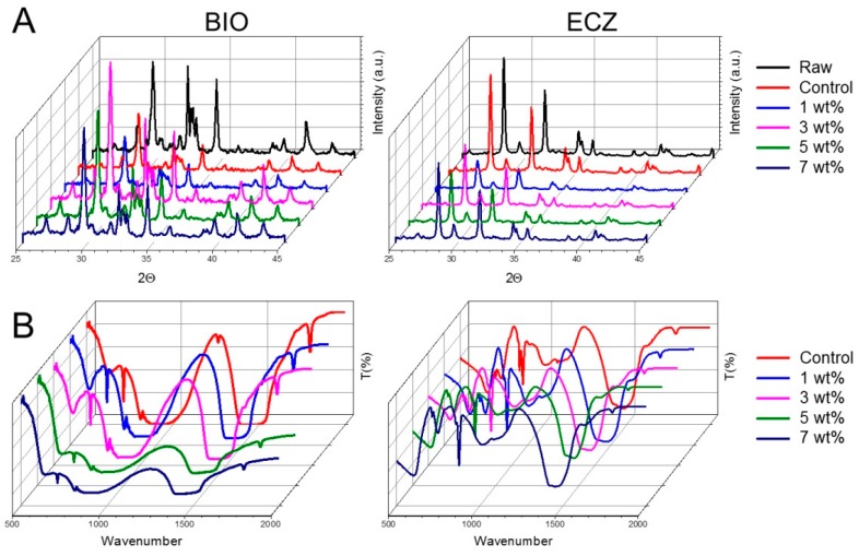
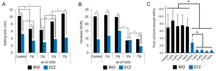
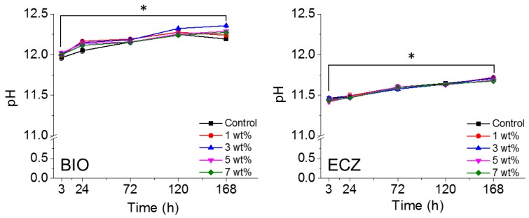
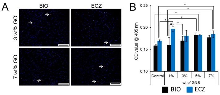
Bioactive calcium silicate cements are widely used to induce mineralization, to cement prosthetic parts, in the management of tooth perforations, and other areas. Nonetheless, they can present clinical disadvantages, such as long setting time and modest physico-mechanical properties. The objective of this work was to evaluate the potential of graphene nanosheets (GNS) to improve two bioactive cements. GNS were obtained via reduction of graphite oxide. GNS were mixed (1, 3, 5, and 7 wt %) with Biodentine (BIO) and Endocem Zr (ECZ), and the effects on setting time, hardness, push-out strength, pH profile, cell proliferation, and mineralization were evaluated. Statistics were performed with two-way ANOVA and Tukey test (α = 0.05). GNS has not interfered in the composition of the set cements as confirmed by Raman, FT-IR and XRD. GNS (1 and 3 wt %) shortened the setting time, increased hardness of both materials but decreased significantly the push-out strength of ECZ. pH was not affected but 1 wt % and 7 wt % to ECZ and 5 wt % to BIO increased the mineralization compared to the controls. In summary, GNS may be an alternative to improve the physico-mechanical properties and bioactivity of cements. Nonetheless, the use of GNS may not be advised for all materials when effective bonding is a concern.

Keywords: Biodentine; dental pulp stem cells; graphene; mineral trioxide aggregate; push-out bond strength.


Conflict of interest statement

Authors declare no conflict of interest. The funding sponsors had no role in the design of the study; in the collection, analyses, or interpretation of data; in the writing of the manuscript, and in the decision to publish the results.

Figures

Figure 1
Figure 1
GNS characterization via SEM (A), scale bar = 10 µm; Raman (B); and FT-IR (C) spectroscopy.
Figure 2
Figure 2
SEM of fracture surface of specimens showed that GNS (arrows) was evenly dispersed in the cements (scale bar = 50 µm).
Figure 3
Figure 3
Raman spectroscopy shows the presence of peaks at 1583 and 2700 cm−1 for all of the experimental groups.
Figure 4
Figure 4
XRD (A) and FT-IR (B) for all conditions tested (Raw = unmodified powder before mixing).
Figure 5
Figure 5
Setting time (A); hardness (B); and push-out bond strength (C) of the groups tested (* represents statistical significance between groups, p < 0.05). The fracture modes obtained in the push-out test are available in Figure S1.
Figure 6
Figure 6
pH variation of both materials with different wt % of GNS. The addition of GNS did not change the alkaline potential of the materials tested (* represents statistical significance between groups, p < 0.05).
Figure 7
Figure 7
Cell viability was not compromised by the addition of 3 and 7 wt % of GNS after five days. The arrow indicates the dead cell in culture (A); bioactivity was assessed by Alizarin red S staining. There was no statistical difference for BIO and ECZ for the same condition except for 1 wt % of GNS (B). Scale bar = 20 µm, the full set of data for three and five days for cell viability is available in Figure S2.

Similar articles

Cited by

References

    1. Caron G., Azerad J., Faure M.O., Machtou P., Boucher Y. Use of a new retrograde filling material (biodentine) for endodontic surgery: Two case reports. Int. J. Oral Sci. 2014;6:250–253. doi: 10.1038/ijos.2014.25. - DOI - PMC - PubMed
    1. Vidal K., Martin G., Lozano O., Salas M., Trigueros J., Aguilar G. Apical closure in apexification: A review and case report of apexification treatment of an immature permanent tooth with biodentine. J. Endod. 2016;42:730–734. doi: 10.1016/j.joen.2016.02.007. - DOI - PubMed
    1. Michel A., Erber R., Frese C., Gehrig H., Saure D., Mente J. In vitro evaluation of different dental materials used for the treatment of extensive cervical root defects using human periodontal cells. Clin. Oral Investig. 2016;21:753–761. doi: 10.1007/s00784-016-1830-3. - DOI - PubMed
    1. Marconyak L.J., Kirkpatrick T.C., Roberts H.W., Roberts M.D., Aparicio A., Himel V.T., Sabey K.A. A comparison of coronal tooth discoloration elicited by various endodontic reparative materials. J. Endod. 2016;42:470–473. doi: 10.1016/j.joen.2015.10.013. - DOI - PubMed
    1. Kang S.-H., Shin Y.-S., Lee H.-S., Kim S.-O., Shin Y., Jung I.-Y., Song J.S. Color changes of teeth after treatment with various mineral trioxide aggregate–based materials: An ex vivo study. J. Endod. 2015;41:737–741. doi: 10.1016/j.joen.2015.01.019. - DOI - PubMed

LinkOut - more resources