Review
. 2017 Jul 31;10(8):884.
doi: 10.3390/ma10080884.

Metallic Biomaterials: Current Challenges and Opportunities

Affiliations
Review

Metallic Biomaterials: Current Challenges and Opportunities

Karthika Prasad et al. Materials (Basel). .

Abstract

Metallic biomaterials are engineered systems designed to provide internal support to biological tissues and they are being used largely in joint replacements, dental implants, orthopaedic fixations and stents. Higher biomaterial usage is associated with an increased incidence of implant-related complications due to poor implant integration, inflammation, mechanical instability, necrosis and infections, and associated prolonged patient care, pain and loss of function. In this review, we will briefly explore major representatives of metallic biomaterials along with the key existing and emerging strategies for surface and bulk modification used to improve biointegration, mechanical strength and flexibility of biometals, and discuss their compatibility with the concept of 3D printing.

Keywords: advanced materials; biomaterial; implant; inflammation; surface modification.


Conflict of interest statement

The authors declare no conflict of interest.

Figures

Figure 1
Figure 1
(a) Corrosion rates of Mg-based alloys in physiologically relevant solutions [12]; (b) Images of Mg95–xZnxCa5 (at %) implanted in rat femurs, reconstructed from in vivo μ-CT scans. Note visible degradation after 30 days for the x = 28 sample, while minimal and no degradation are visible on the x = 32 and x = 35 samples, respectively [13]; (c) Radiographs of mice distal femora with and without implanted high-entropy CaMgZnSrYb alloy, immediately after implantation, and 4 weeks postoperatively. The sample shows no gas formation, no inflammation, and enhanced circumferential osteogenesis in the implanted bone (yellow arrow), indicating new bone formation [14]. Reproduced with permission from [14,15].
Figure 2
Figure 2
Top image: Photographs of a patient with erythema and rash after implantation of a stainless steel bar. Bottom image: typical symptoms reported in patients implanted with steel bars as part of a Nuss procedure for repair of pectus excavatum. Patients did not have a history of metal allergy prior to implantation. Reproduced with permission from [52].
Figure 3
Figure 3
(a) Stent to be inserted in artery to keep the passageway open. Commonly made of stainless steel; (b) Prototype of biodegradable stent; (top) as fabricated; (mid) crimped onto a balloon catheter, and (bottom) expanded to 3 mm by 6 atm of pressure. Reproduced with permission from [55,56].
Figure 4
Figure 4
Histological observation of hard tissue in contact with porous tantalum implants at week (A) 2; (B) 4; (C) 8 and (D) 12 after implantation (methylene blue staining). Reproduced with permission from [95].
Figure 5
Figure 5
Degradation of magnesium staples under in vitro and in vivo conditions. Optical visualisation of morphology of Mg staples after immersion in simulated body fluid at pH of 4 for (a) 3; (b) 7; (c) 11; and (d) 14 days. Photographs of Mg staples implanted into an animal model for (e) 7 and (f) 90 days; and (g) Ti staples implanted for 90 days. Reproduced with permission from [116].
Figure 6
Figure 6
Fluorescence images of initial adherent bone marrow mesenchymal stem cells (BMSCs) stained with DAPI after 1 h (a) and cell numbers measured by counting cells for 0.5 h, 1 h, and 2 h (b). Cell attachment on NT10 (titanium nanotube with 10 nm diameter) was significantly improved compared with control Ti surfaces for 1 h and 2 h. In contrast, cell attachment was inhibited on NT30 (titanium nanotube with 30 nm diameter) and NT60 (titanium nanotube with 60 nm diameter) at each time interval adopted in this study. All data were expressed as the mean ± standard deviation (SD) and ‘p’, the level of significance. p < 0.05 was considered significant, and p < 0.01 was considered highly significant. Here, the mean ± SD N = 3, and * p < 0.05, and ** p < 0.01 compared with the T; # p < 0.05 and ## p < 0.01 compared with the NT10; & p < 0.05 and && p < 0.01 compared with NT30. Reproduced with permission from [124].
Figure 7
Figure 7
Fluorescent microscopy images of endothelial cell proliferation on nanostructured Ti compared to conventional Ti. Reproduced with permission from [129].
Figure 8
Figure 8
Wear morphology of two differently-processed Ti-29Nb-13Ta-4.6Zr (TNZT) alloys (a) TNZT1 and (b) TNZT3 and (c) a Ti-6Al-4V alloy (TAV1) sliding on a stainless steel plate; (d) TNZT1; (e) TNZT3; and (f) TAV1 sliding on ultra-high-molecular-weight polyethylene (UHMWPE); (g) TNZT1; (h) TNZT3; and (i) TAV1 sliding on a pig bone in 0.9% NaCl. Reproduced with permission from [81].
Figure 9
Figure 9
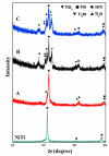
Low angle XRD phase analysis of NiTi surface implanted by nitrogen ions: A, B and C indicated the intensities at different concentration of nitrogen, such as 6.9 × 10¹⁷ ion cm−2 (sample A) 1.4 × 10¹⁸ ion cm−2 (sample B), and 1.8 × 10¹⁸ ion cm−2 (sample C). Reproduced with permission from [161].
Figure 10
Figure 10
SEM images showing the corrosion after 3, 15 and 30 days. The image shows the increased corrosion rate of the Zn- implanted iron (b,d,f) in comparison to the pure iron (a,c,e) samples, with pure Fe samples degrading mostly along the grain boundaries (e). Reproduced with permission from [164].
Figure 11
Figure 11
Grain structures or morphologies of magnesium alloy (AZ31) (series 1), iron alloy (Fe-Mn-1Ca) (series 2) and commercially pure (CP) titanium (series 3). Series 1 depicts the cyclic extrusion compression (CEC) process of AZ31 at 300 °C, (1a) showing the alloy “as extruded’, (1b) showing the alloy after 7 passes and (1c) showing the alloy after 15 passes. Series 2 shows the morphology of the iron based alloy before (2a) and after (2b) the binder-jet-3D printing process has been completed. Series 3 shows the morphology of a CP titanium sample after 3 laser shock peening impacts. (3a) shows the morphology of the sample close to the surface, while (3b) shows the morphology of the sample less than 0.5 mm below the surface and (3c) shows the sample morphology at about 2 mm below the surface. All images—series 1, 2 & 3—were reproduced with permission from [166], [4] and [3], respectively.

References

    1. Global Bio-Implants Market Worth $134.3 Billion by 2017. [(accessed on 19 June 2017)];2017 Available online: http://www.marketsandmarkets.com/PressReleases/bio-implants.asp.
    1. Ige O.O., Umoru L.E., Aribo S. Natural products: A minefield of biomaterials. ISRN Mater. Sci. 2012;2012:983062:1–983062:20. doi: 10.5402/2012/983062. - DOI
    1. Lu J.Z., Wu L.J., Sun G.F., Luo K.Y., Zhang Y.K., Cai J., Cui C.Y., Luo X.M. Microstructural response and grain refinement mechanism of commercially pure titanium subjected to multiple laser shock peening impacts. Acta Mater. 2017;127:252–266. doi: 10.1016/j.actamat.2017.01.050. - DOI
    1. Hong D., Chou D.-T., Velikokhatnyi O.I., Roy A., Lee B., Swink I., Issaev I., Kuhn H.A., Kumta P.N. Binder-jetting 3D printing and alloy development of new biodegradable Fe-Mn-Ca/Mg alloys. Acta Biomater. 2016;45:375–386. doi: 10.1016/j.actbio.2016.08.032. - DOI - PubMed
    1. Trivedi P., Nune K.C., Misra R.D.K., Goel S., Jayganthan R., Srinivasan A. Grain refinement to submicron regime in multiaxial forged Mg-2Zn-2Gd alloy and relationship to mechanical properties. Mater. Sci. Eng. A. 2016;668:59–65. doi: 10.1016/j.msea.2016.05.050. - DOI

LinkOut - more resources