. 2016 Mar 23;9(4):221.
doi: 10.3390/ma9040221.

Aqueous-Based Coaxial Electrospinning of Genetically Engineered Silk Elastin Core-Shell Nanofibers

Affiliations

Aqueous-Based Coaxial Electrospinning of Genetically Engineered Silk Elastin Core-Shell Nanofibers

Jingxin Zhu et al. Materials (Basel). .

Abstract

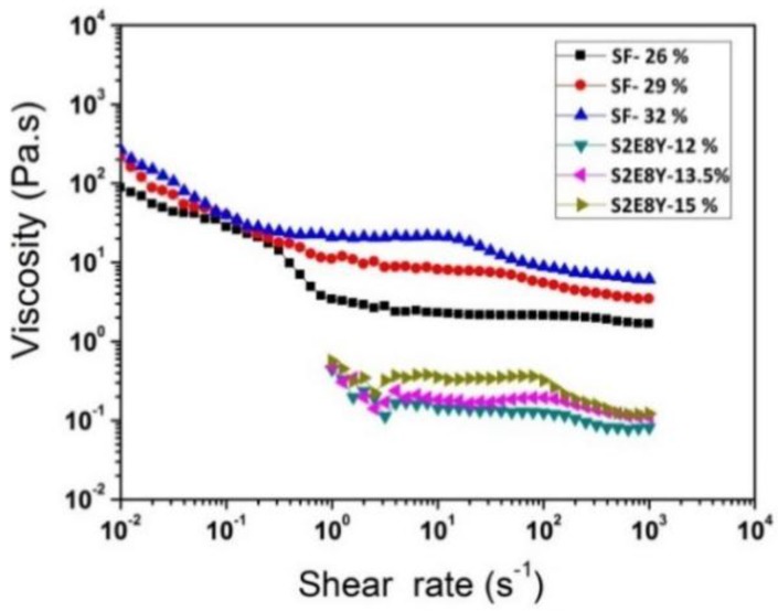
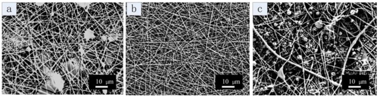
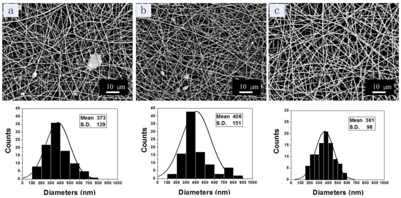
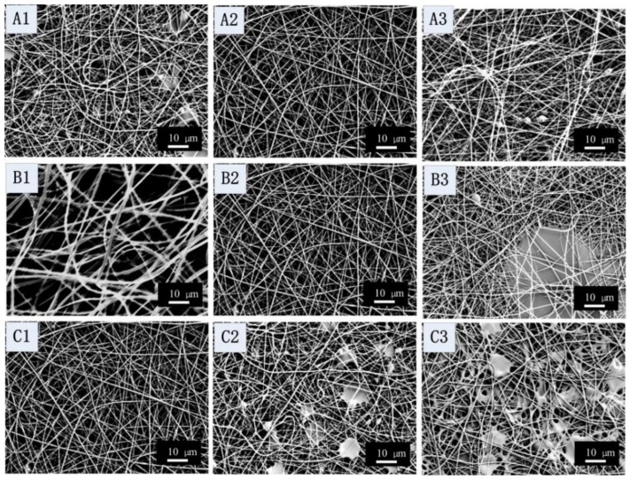
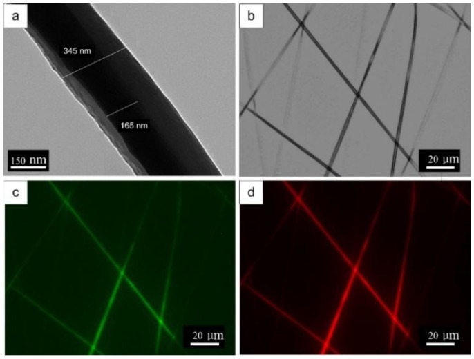
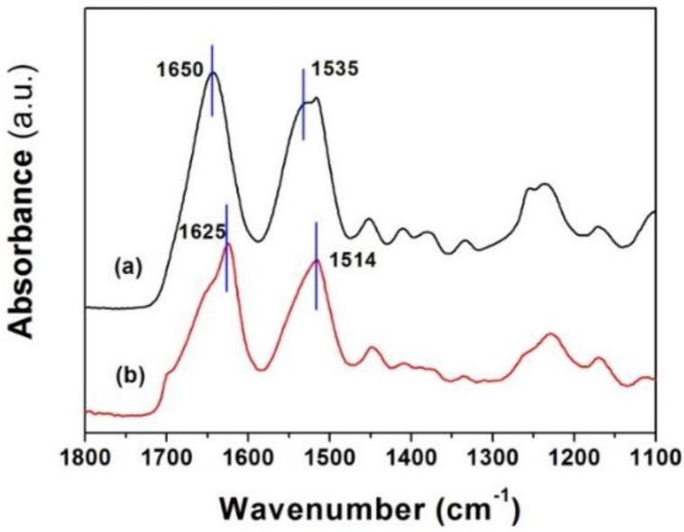
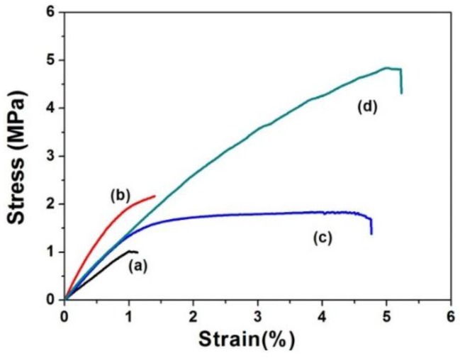
A nanofabrication method for the production of flexible core-shell structured silk elastin nanofibers is presented, based on an all-aqueous coaxial electrospinning process. In this process, silk fibroin (SF) and silk-elastin-like protein polymer (SELP), both in aqueous solution, with high and low viscosity, respectively, were used as the inner (core) and outer (shell) layers of the nanofibers. The electrospinnable SF core solution served as a spinning aid for the nonelectrospinnable SELP shell solution. Uniform nanofibers with average diameter from 301 ± 108 nm to 408 ± 150 nm were obtained through adjusting the processing parameters. The core-shell structures of the nanofibers were confirmed by fluorescence and electron microscopy. In order to modulate the mechanical properties and provide stability in water, the as-spun SF-SELP nanofiber mats were treated with methanol vapor to induce β-sheet physical crosslinks. FTIR confirmed the conversion of the secondary structure from a random coil to β-sheets after the methanol treatment. Tensile tests of SF-SELP core-shell structured nanofibers showed good flexibility with elongation at break of 5.20% ± 0.57%, compared with SF nanofibers with an elongation at break of 1.38% ± 0.22%. The SF-SELP core-shell structured nanofibers should provide useful options to explore in the field of biomaterials due to the improved flexibility of the fibrous mats and the presence of a dynamic SELP layer on the outer surface.

Keywords: coaxial electrospinning; core-shell structure; silk fibroin; silk-elastin-like protein polymer.


Conflict of interest statement

The authors have no other relevant affiliations or financial involvement with any organization or entity with a financial interest in or financial conflict with the subject matter or materials discussed in the manuscript. This includes employment, consultancies, honoraria, stock ownership or options, expert testimony, grants or patents received or pending or royalties.

Figures

Figure 1
Figure 1
Sodium dodecyl sulfate polyacrylamide gel electrophoresis (SDS-PAGE) of purified S2E8Y and regenerated silk fibroin (SF).
Figure 2
Figure 2
Rheology of SF and S2E8Y solutions with different concentrations.
Figure 3
Figure 3
Scanning electron microscopy (SEM) images of coaxial electrospun SF-SELP nanofibers with various SF concentrations: (a) 26 wt % (b) 29 wt % (c) 32 wt %. The concentration of the shell SELP solution was fixed at 15%. The inner flow rate was 0.4 mL/h, the outer flow rate 0.17 mL/h, the tip to collector distance 12 cm and the applied voltage 20 kV.
Figure 4
Figure 4
SEM images of coaxial electrospun SF-SELP nanofibers with various SELP concentrations: (a) 12 wt % (b) 13.5 wt % (c) 15 wt %. The concentration of SF solution was 29%. The inner flow rate was 0.4 mL/h, the outer flow rate 0.17 mL/h, the tip to collector distance 10 cm and the applied voltage 20 kV.
Figure 5
Figure 5
SEM images of coaxial electrospun SF-SELP nanofibers at different processes parameters: (A1, A2, A3) inner flow rates at 0.2, 0.4, and 0.6 mL/h, respectively; (B1, B2, B3) outer flow rates at 0.07, 0.17, and 0.34 mL/h, respectively; (C1, C2, C3) applied voltage at 20, 25, 30 kV, respectively. Other processing parameters are listed in Table 2.
Figure 6
Figure 6
Conditions where to electrospin uniform SF-SELP nanofibers were generated: (A) mean diameters of fibers (nm) with outer SELP concentration (%) and working distance (cm); (B) mean diameters of fibers with inner flow rates (mL/h) and outer SELP concentration (%).
Figure 7
Figure 7
Core-shell structures of the SF-SELP nanofibers: (a) transmission electron microscopy (TEM) image and (b) phase-contrast image of fibers; (c) fluorescent image showing fluorescein isothiocyanate (FTIC)–dextran stained S2E8Y shell phase; and (d) fluorescent image showing rhodamine B stained SF cores phase.
Figure 8
Figure 8
Fourier transform infrared spectroscopy (FTIR) spectra for SF-SELP core-shell fiber mats before and after methanol vapor treatment: (a) before methanol vapor treatment; and (b) after methanol vapor treatment.
Figure 9
Figure 9
Stress-strain curves of SF fibers mats and SF-SELP core-shell fibers mats before and after methanol treatment: (a) SF fibers mats; (b) SF fibers mats after methanol treatment; (c) SF-SELPs core-sell fiber mats; and (d) SF-SELPs core-sell fiber mats after methanol treatment.

Similar articles

Cited by

References

    1. Bhardwaj N., Kundu S.C. Electrospinning: A fascinating fiber fabrication technique. Biotechnol. Adv. 2010;28:325–347. doi: 10.1016/j.biotechadv.2010.01.004. - DOI - PubMed
    1. Li H., Xu Y., Xu H., Chang J. Electrospun membranes: Control of the structure and structure related applications in tissue regeneration and drug delivery. J. Mater. Chem. B. 2014;2:5492–5510. doi: 10.1039/C4TB00913D. - DOI - PubMed
    1. Alessandrino A., Marelli B., Arosio C., Fare S., Tanzi M.C., Freddi G. Electrospun silk fibroin mats for tissue engineering. Eng. Life Sci. 2008;8:219–225. doi: 10.1002/elsc.200700067. - DOI
    1. Li C.M., Vepari C., Jin H.J., Kim H.J., Kaplan D.L. Electrospun silk-BMP-2 scaffolds for bone tissue engineering. Biomaterials. 2006;27:3115–3124. doi: 10.1016/j.biomaterials.2006.01.022. - DOI - PubMed
    1. Vepari C., Kaplan D.L. Silk as a biomaterial. Prog. Polym. Sci. 2007;32:991–1007. doi: 10.1016/j.progpolymsci.2007.05.013. - DOI - PMC - PubMed

LinkOut - more resources