. 2016 Mar 30;9(4):249.
doi: 10.3390/ma9040249.

Rapid Assay to Assess Bacterial Adhesion on Textiles

Affiliations

Rapid Assay to Assess Bacterial Adhesion on Textiles

Sabrina Schmidt-Emrich et al. Materials (Basel). .

Abstract

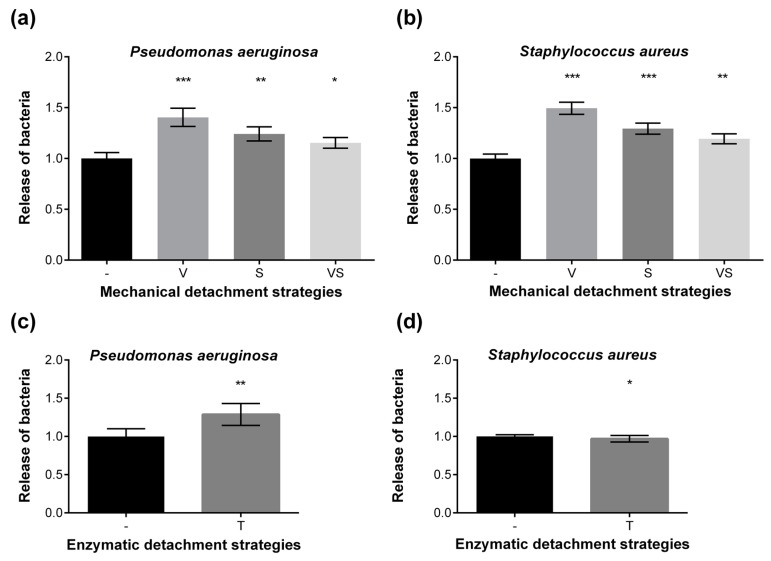
Textiles are frequently colonized by microorganisms leading to undesired consequences like hygienic problems. Biocidal coatings often raise environmental and health concerns, thus sustainable, biocide-free coatings are of interest. To develop novel anti-adhesive textile coatings, a rapid, reliable, and quantitative high-throughput method to study microbial attachment to fabrics is required, however currently not available. Here, a fast and reliable 96-well plate-based screening method is developed. The quantification of bacterial adhesion is based on nucleic acid staining by SYTO9, with Pseudomonas aeruginosa and Staphylococcus aureus as the model microorganisms. Subsequently, 38 commercially available and novel coatings were evaluated for their anti-bacterial adhesion properties. A poly(l-lysine)-g-poly(ethylene glycol) coating on polyester textile substratum revealed an 80% reduction of bacterial adhesion. Both the coating itself and the anti-adhesive property were stable after 20 washing cycles, confirmed by X-ray analysis. The assay provides an efficient tool to rapidly screen for non-biocidal coatings reducing bacterial attachment.

Keywords: antifouling; bacterial adhesion; biofilm; microtiter plate; textile coating.


Conflict of interest statement

The authors declare no conflict of interest.

Figures

Figure A1
Figure A1
Calibration curves of cell count (logCFU/mL) and relative fluorescence intensity (logRFU) for P. aeruginosa (upper panel) and S. aureus (lower panel).
Figure 1
Figure 1
Work flow of microplate screening to quantify bacterial attachment to textile coatings. SYTO9 staining was applied for the large screening, alternative techniques such as bioluminescence assay and viable cell count were tested as well.
Figure 2
Figure 2
SEM image of P. aeruginosa cells on polyester fiber after 2 h attachment.
Figure 3
Figure 3
Mechanical (a,b) and enzymatic (c,d) detachment of P. aeruginosa (a,c) and S. aureus (b,d) cells from textiles. Comparison of mechanical (-: no treatment, V: vortexing, S: sonication, VS: vortexing plus sonication) and enzymatic (-: no enzymes, T: trypsin) strategies to dislodge attached bacterial cells from cotton. The release of bacteria cells was calculated for each dislodgement technique by comparing untreated controls (no mechanical or enzymatic detachment) which were set as 1.0. The experiment was repeated twice independently with a sample number of n = 31 per condition for mechanical treatment, and n = 16 for trypsin treatment. Columns are displayed as means, error bars are shown as plus/minus standard error of the mean. Statistical analyses were performed via the unpaired, parametric, two-tailed Student’s t-test. *** p < 0.001; ** p < 0.01; * p ≤ 0.05 vs. no treatment/no enzymes.
Figure 4
Figure 4
Screening of anti-adhesive coatings using the microplate assay. A number of 38 textile coatings on either cotton or polyester textiles were tested for their anti-adhesive properties against the bacterial attachment. The most-promising coatings against P. aeruginosa (a) and S. aureus (b) are shown. Each coated sample type was compared to its uncoated counterpart. The difference between the bacterial adhesion to coated and non-coated fabrics (set as 100%) is displayed as reduction of attached bacteria in percentage. All experiments were repeated twice independently with a sample number of n = 8 per condition. Statistical analyses were performed via the unpaired, parametric, two-tailed Student’s t-test. *** p < 0.001; ** p < 0.01; * p < 0.05; (c) shows the washing resistance of anti-adhesive textile coatings with Coating C derivative. Samples were washed 5 and 20 times, respectively, and tested afterwards for the reduction of P. aeruginosa adhesion (%) using the microplate assay. This experiment was conducted twice independently. The reduction of bacterial attachment kept at more than 80% after 5 and 20 times washing.
Figure 5
Figure 5
Comparison of high-resolution elemental C1s photoelectron spectra. (a): uncoated PET textile substratum; (b,c): different positions of PLL-g-PEG coated PET textile sample. The bold black line represents the experimental spectrum, the dashed lines are the bands from the curve fitting with corresponding assignments as C–C/C–H (285.0 eV), C–O/C–N (286.4 eV), N–C=O (288.1 eV), and O–C=O (289.0 eV).

Similar articles

Cited by

References

    1. Szostak-Kotowa J. Biodeterioration of textiles. Int. Biodeterior. Biodegrad. 2004;53:165–170. doi: 10.1016/S0964-8305(03)00090-8. - DOI
    1. Bajpai V., Bajpai S., Jha M.K., Dey A., Ghosh S. Microbial adherence on textile materials: A review. J. Environ. Res. Dev. 2011;5:666–672.
    1. Gao Y., Cranston R. Recent advances in antimicrobial treatments of textiles. Text. Res. J. 2008;78:60–72.
    1. Gouveia I.C. Nanobiotechnology: A new strategy to develop non-toxic antimicrobial textiles for healthcare applications. J. Biotechnol. 2010;150:349. doi: 10.1016/j.jbiotec.2010.09.387. - DOI
    1. Silver S. Bacterial silver resistance: Molecular biology and uses and misuses of silver compounds. FEMS Microbiol. Rev. 2003;27:341–353. doi: 10.1016/S0168-6445(03)00047-0. - DOI - PubMed

LinkOut - more resources