. 2016 Apr 7;9(4):276.
doi: 10.3390/ma9040276.

In Vitro Testing of Scaffolds for Mesenchymal Stem Cell-Based Meniscus Tissue Engineering-Introducing a New Biocompatibility Scoring System

Affiliations

In Vitro Testing of Scaffolds for Mesenchymal Stem Cell-Based Meniscus Tissue Engineering-Introducing a New Biocompatibility Scoring System

Felix P Achatz et al. Materials (Basel). .

Abstract

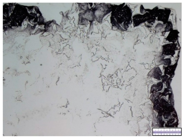
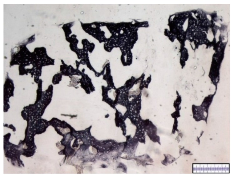
A combination of mesenchymal stem cells (MSCs) and scaffolds seems to be a promising approach for meniscus repair. To facilitate the search for an appropriate scaffold material a reliable and objective in vitro testing system is essential. This paper introduces a new scoring for this purpose and analyzes a hyaluronic acid (HA) gelatin composite scaffold and a polyurethane scaffold in combination with MSCs for tissue engineering of meniscus. The pore quality and interconnectivity of pores of a HA gelatin composite scaffold and a polyurethane scaffold were analyzed by surface photography and Berliner-Blau-BSA-solution vacuum filling. Further the two scaffold materials were vacuum-filled with human MSCs and analyzed by histology and immunohistochemistry after 21 days in chondrogenic media to determine cell distribution and cell survival as well as proteoglycan production, collagen type I and II content. The polyurethane scaffold showed better results than the hyaluronic acid gelatin composite scaffold, with signs of central necrosis in the HA gelatin composite scaffolds. The polyurethane scaffold showed good porosity, excellent pore interconnectivity, good cell distribution and cell survival, as well as an extensive content of proteoglycans and collagen type II. The polyurethane scaffold seems to be a promising biomaterial for a mesenchymal stem cell-based tissue engineering approach for meniscal repair. The new score could be applied as a new standard for in vitro scaffold testing.

Keywords: biocompatibility; chondrogenesis; collagen; composite scaffold; gelatin; human mesenchymal stem cells; hyaluronic acid; meniscus; polyurethane scaffold.


Conflict of interest statement

The authors declare no conflict of interest. Parts of the project (Actifit® biomaterial supply) were supported by Orteq. The funding sponsors had no role in the design of study; in the collection, analysis or interpretation of data; in the writing of the manuscript, and in the decision to publish the results.

Figures

Figure 1
Figure 1
Macroscopic enhanced image of a hyaluronic acid gelatin composite scaffold. Scale bar = 1 mm.
Figure 2
Figure 2
Macroscopic enhanced image of an Actifit® scaffold. Scale bar = 1 mm.
Figure 3
Figure 3
Illustration of pore interconnectivity in a hyaluronic acid gelatin composite scaffold. Black areas represent accessible pores whereas white areas signify secluded pores. Arrow A points to an interconnected, accessible pore. Arrow B points to a non-interconnected, secluded pore. Most of the scaffold’s area is shows a black color, thus proving excellent pore interconnectivity. Magnification bar = 1 mm.
Figure 4
Figure 4
Illustration of pore interconnectivity in an Actifit® scaffold. Black areas represent accessible pores whereas white areas signify secluded pores. A major part of the scaffold’s area is shows a black color, thus proving good pore interconnectivity Magnification bar = 1 mm.
Figure 5
Figure 5
Representative histological slide of a hyaluronic acid gelatin composite scaffold after 21 days of in vitro chondrogenesis with visible central necrosis. DMMB staining. Proteoglycan-rich extracellular matrix appears red, scaffold parts appear blue. Magnification bar = 500 μm.
Figure 6
Figure 6
Representative histological slide of an Actifit® scaffold after 21 days of in vitro chondrogenesis. DMMB staining. Proteoglycan-rich extracellular matrix appears red, scaffold parts appear grey and porous. Magnification bar = 500 μm.
Figure 7
Figure 7
Representative slide of a hyaluronic acid gelatin composite scaffold after 21 days of in vitro chondrogenesis. Collagen type I immunohistochemistry. Collagen type I-rich areas appear black. Magnification bar = 500 μm.
Figure 8
Figure 8
Representative slide of an Actifit® scaffold after 21 days of in vitro chondrogenesis. Collagen type I immunohistochemistry. Collagen type I-rich areas appear black. Magnification bar = 500 μm.
Figure 9
Figure 9
Representative slide of a hyaluronic acid gelatin composite scaffold after 21 days of in vitro chondrogenesis. Collagen type II immunohistochemistry. Collagen type II-rich areas appear black. Magnification bar = 500 μm.
Figure 10
Figure 10
Representative slide of an Actifit® scaffold after 21 days of in vitro chondrogenesis. Collagen type II immunohistochemistry. Collagen type II-rich areas appear black. Magnification bar = 500 μm.
Figure 11
Figure 11
Overall Scoring Results of the different scaffolds. Error bars indicate 95% confidence interval.
Figure 12
Figure 12
Average scoring results of the different scaffolds for single score item viability of cells. Error bars indicating 95% confidence interval. Scoring item was assessed in a semi-quantitative manner. Cell viability was statistically significant better (Mann–Whitney-U test, ** = p < 0.05) in the Actifit® scaffolds compared to the HA (hyaluronic acid) gelatin composite scaffolds.

Similar articles

Cited by

References

    1. Verdonk P., Beaufils P., Bellemans J., Djian P., Heinrichs E.-L., Huysse W., Laprell H., Siebold R., Verdonk R., Colombet P., et al. Successful Treatment of Painful Irreparable Partial Meniscal Defects with a Polyurethane Scaffold: Two-Year Safety and Clinical Outcomes. Am. J. Sports Med. 2012;40:844–853. doi: 10.1177/0363546511433032. - DOI - PubMed
    1. Brindle T., Nyland J., Johnson D.L. The meniscus: Review of basic principles with application to surgery and rehabilitation. J. Athl. Train. 2001;36:160–169. - PMC - PubMed
    1. Petty C.A., Lubowitz J.H. Does arthroscopic partial meniscectomy result in knee osteoarthritis? A systematic review with a minimum of 8 years’ follow-up. Arthroscopy. 2011;27:419–424. doi: 10.1016/j.arthro.2010.08.016. - DOI - PubMed
    1. Liu C., Toma I.C., Mastrogiacomo M., Krettek C., Lewinski G., von Jagodzinski M. Meniscus reconstruction: Today’s achievements and premises for the future. Arch. Orthop. Trauma. Surg. 2013;133:95–109. doi: 10.1007/s00402-012-1624-2. - DOI - PubMed
    1. Levy I.M., Torzilli P.A., Gould J.D., Warren R.F. The effect of lateral meniscectomy on motion of the knee. J. Bone Joint Surg. Am. 1989;71:401–406. - PubMed

LinkOut - more resources