. 2016 Apr 14;9(4):285.
doi: 10.3390/ma9040285.

A Hydrogel Model Incorporating 3D-Plotted Hydroxyapatite for Osteochondral Tissue Engineering

Affiliations

A Hydrogel Model Incorporating 3D-Plotted Hydroxyapatite for Osteochondral Tissue Engineering

Michal Bartnikowski et al. Materials (Basel). .

Abstract

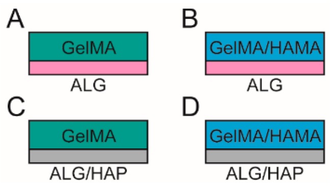
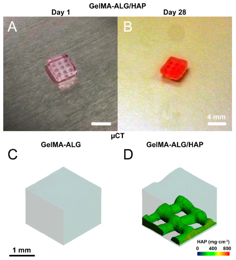
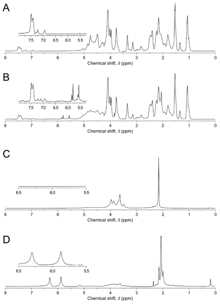
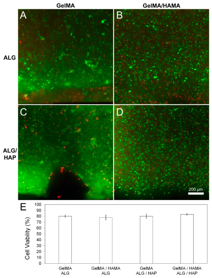
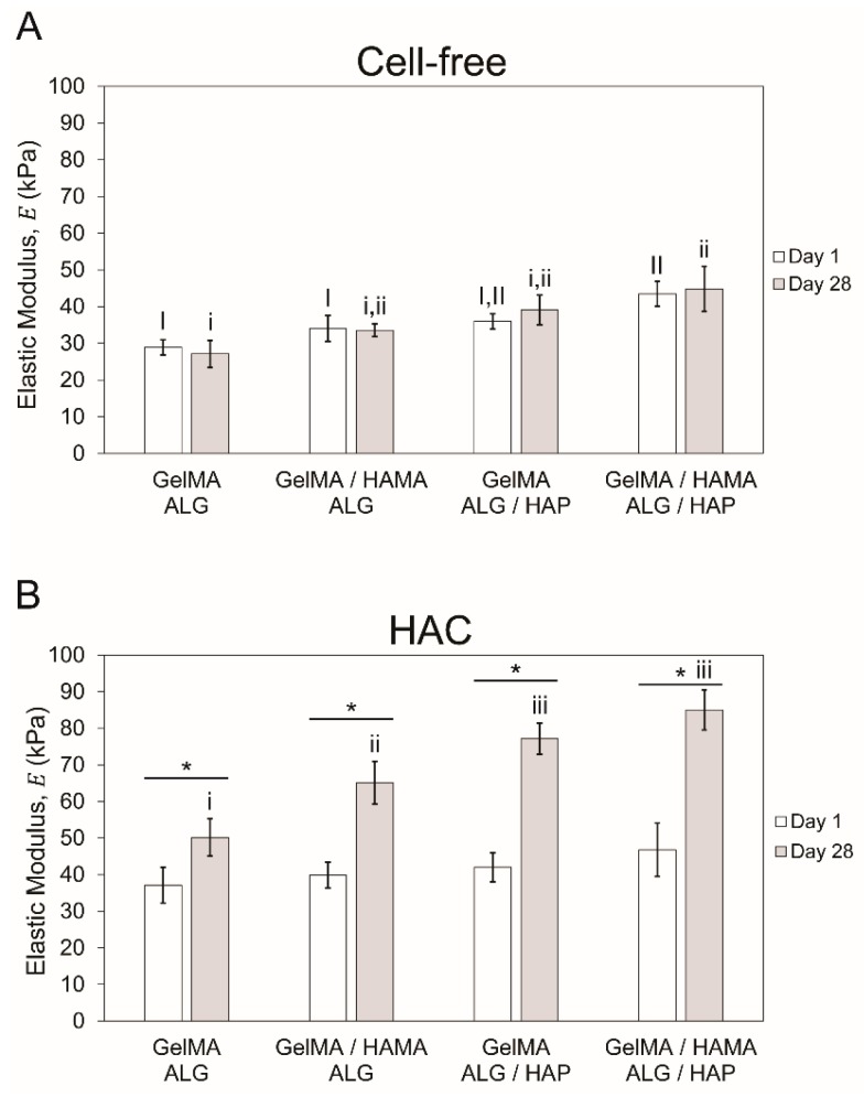
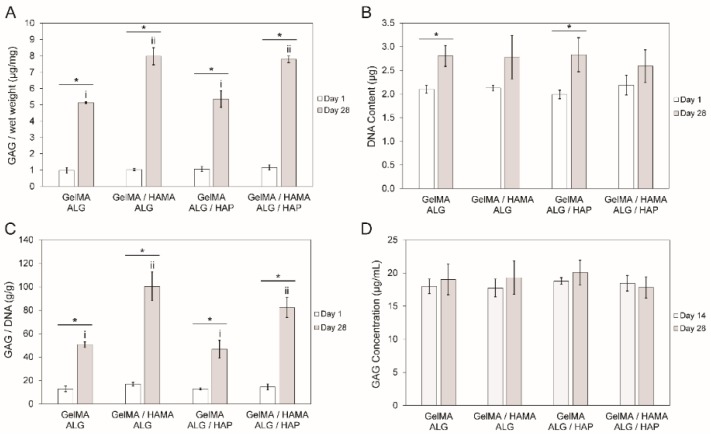
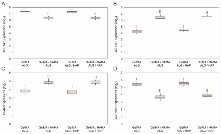
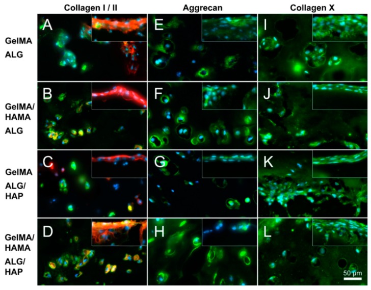
The concept of biphasic or multi-layered compound scaffolds has been explored within numerous studies in the context of cartilage and osteochondral regeneration. To date, no system has been identified that stands out in terms of superior chondrogenesis, osteogenesis or the formation of a zone of calcified cartilage (ZCC). Herein we present a 3D plotted scaffold, comprising an alginate and hydroxyapatite paste, cast within a photocrosslinkable hydrogel made of gelatin methacrylamide (GelMA), or GelMA with hyaluronic acid methacrylate (HAMA). We hypothesized that this combination of 3D plotting and hydrogel crosslinking would form a high fidelity, cell supporting structure that would allow localization of hydroxyapatite to the deepest regions of the structure whilst taking advantage of hydrogel photocrosslinking. We assessed this preliminary design in terms of chondrogenesis in culture with human articular chondrocytes, and verified whether the inclusion of hydroxyapatite in the form presented had any influence on the formation of the ZCC. Whilst the inclusion of HAMA resulted in a better chondrogenic outcome, the effect of HAP was limited. We overall demonstrated that formation of such compound structures is possible, providing a foundation for future work. The development of cohesive biphasic systems is highly relevant for current and future cartilage tissue engineering.

Keywords: 3D plotting; alginate; cartilage tissue engineering; chondrocyte; chondrogenesis; gelatin; hydrogel; hydroxyapatite.


Conflict of interest statement

The authors declare no conflict of interest.

Figures

Figure 1
Figure 1
Schematic illustration of the groups included in the study.Groups used in the study comprised (A): gelatin methacrylamide (GelMA) with an alginate (ALG) paste scaffold; (B): GelMA with hyaluronic acid methacrylate (HAMA) with an ALG paste scaffold; (C): GelMA with a combined alginate and hydroxyapatite paste (ALG/HAP) scaffold; and (D): GelMA/HAMA with an ALG/HAP scaffold.
Figure 2
Figure 2
Images of hydrogel/scaffold hybrid constructs. (A) photo illustrating ALG/HAP scaffolds crosslinked within GelMA hydrogels on day 1 and (B) day 28. Scale bar 4 mm; (C,D) scaffolds imaged using micro-computed tomography (µCT), with thresholding used to isolate high density components (colored scale illustrates mg·cm−3 of HAP); (C) GelMA-ALG and (D) GelMA-ALG/HAP illustrated, scale bar 1 mm as indicated.
Figure 3
Figure 3
Proton Nuclear Magnetic Resonance (1H NMR) spectra of hydrogels. (A): gelatin; (B): gelatin methacrylamide (GelMA); (C): hyaluronic acid (HA); (D): hyaluronic acid methacrylate (HAMA). Areas of interest are inset within each frame. Aromatic peaks in gelatin/GelMA are present at ~chemical shift (δ) 7.4 ppm, with the two free protons on the methacrylate groups present at δ 5.6 and δ 5.8 ppm respectively. In HA, the peak from the n-acetyl protons is present at ~δ 2.1 ppm, with the methacrylate protons used for degree of functionalization (DOF) measurements present in HAMA at δ 5.8 and δ 6.3 ppm.
Figure 4
Figure 4
Cell viability in hydrogel groups on day 1 of culture. Images and quantitated data of cell viability measured using FDA/PI assay on day 1 of culture. Quantification and image analysis was performed using ImageJ. (A): GelMA-ALG; (B): GelMA/HAMA-ALG; (C): GelMA/ALG-HAP; (D): GelMA-HAMA/ALG-HAP; (E): quantified viability from n = 3 samples per group. Scale bar 200 µm.
Figure 5
Figure 5
Mechanical testing results showing compressive elastic modulus. Elastic modulus of samples on days 1 and 28 of culture (n = 3 per group) with (A): cell-free and (B): cell-containing gels shown separately. The four groups in each time point were compared separately to the second time point, with upper case Roman numerals used for day 1 and lower case for day 28. When groups share Roman numerals they are statistically similar. Bars with stars indicate t-test comparison results between one group across two time points.
Figure 6
Figure 6
DNA and GAG content in constructs over the culture period. (A): comparison of day 1 and day 28 results of GAG content (µg) per gel wet weight (mg); (B): DNA content (µg) per gel; (C): GAG content (g) per DNA content (g) in gels and (D): GAG concentration in media (µg·mL−1) on days 14 and 28 of culture. The four groups in each time point were compared separately to the second time point, with upper case Roman numerals used for day 1 and lower case for day 28. When groups share Roman numerals they are statistically similar. Bars with stars indicate t-test comparison results between one group across two time points. All groups have n = 3 samples. In all cases cell-free constructs were used to correct for any material-based content potentially interfering with the GAG measurements.
Figure 7
Figure 7
Gene expression data for the four groups from day 28 samples. The four genes that were analyzed were (A): COL1A1; (B): COL2A1; (C): ACAN and (D): COL10A1 on day 28 of cell culture shown as 2−ΔCt value (after log2 conversion). Statistical analysis was performed on log2 data with each group containing n = 3 samples. A Dunnet’s T3 post-hoc test was used to compare means, with p < 0.05 taken as significant. Shared Roman numerals indicate statistical similarity between gel groups.
Figure 8
Figure 8
Immunofluorescence images illustrating deep and surface sections. Staining of collagen I (red), collagen II (green), aggrecan (green) and collagen X (green) was performed on constructs from all groups on day 28 (as indicated; DAPI nuclei staining in light blue); (A,E,I): GelMA-ALG; (B,F,J): GelMA/HAMA-ALG; (C,G,K): GelMA-ALG/HAP and (D,H,L): GelMA/HAMA-ALG/HAP. Images were taken from the deep hydrogel zone near to the ALG or ALG/HAP scaffold, with surface images inset; scale bar 50 µm.

Similar articles

Cited by

References

    1. Eyre D. Articular cartilage and changes in arthritis: Collagen of articular cartilage. Arthritis Res. 2002;4:30–35. doi: 10.1186/ar380. - DOI - PMC - PubMed
    1. Zhang Y., Wang F., Tan H., Chen G., Guo L., Yang L. Analysis of the mineral composition of the human calcified cartilage zone. Int. J. Med. Sci. 2012;9:353–360. doi: 10.7150/ijms.4276. - DOI - PMC - PubMed
    1. Athanasiou K.A., Darling E.M., DuRaine G.D., Hu J.C., Reddi A.H. Articular Cartilage. CRC Press, Taylor & Francis Group; Boca Raton, FL, USA: 2013. p. 425.
    1. Varghese S., Hwang N.S., Canver A.C., Theprungsirikul P., Lin D.W., Elisseeff J. Chondroitin sulfate based niches for chondrogenic differentiation of mesenchymal stem cells. Matrix Biol. 2008;27:12–21. doi: 10.1016/j.matbio.2007.07.002. - DOI - PubMed
    1. Klein T.J., Rizzi S.C., Reichert J.C., Georgi N., Malda J., Schuurman W., Crawford R.W., Hutmacher D.W. Strategies for zonal cartilage repair using hydrogels. Macromol. Biosci. 2009;9:1049–1058. doi: 10.1002/mabi.200900176. - DOI - PubMed

LinkOut - more resources