. 2016 Jun 29;9(7):532.
doi: 10.3390/ma9070532.

Biomimetic Multispiked Connecting Ti-Alloy Scaffold Prototype for Entirely-Cementless Resurfacing Arthroplasty Endoprostheses-Exemplary Results of Implantation of the Ca-P Surface-Modified Scaffold Prototypes in Animal Model and Osteoblast Culture Evaluation

Affiliations

Biomimetic Multispiked Connecting Ti-Alloy Scaffold Prototype for Entirely-Cementless Resurfacing Arthroplasty Endoprostheses-Exemplary Results of Implantation of the Ca-P Surface-Modified Scaffold Prototypes in Animal Model and Osteoblast Culture Evaluation

Ryszard Uklejewski et al. Materials (Basel). .

Abstract

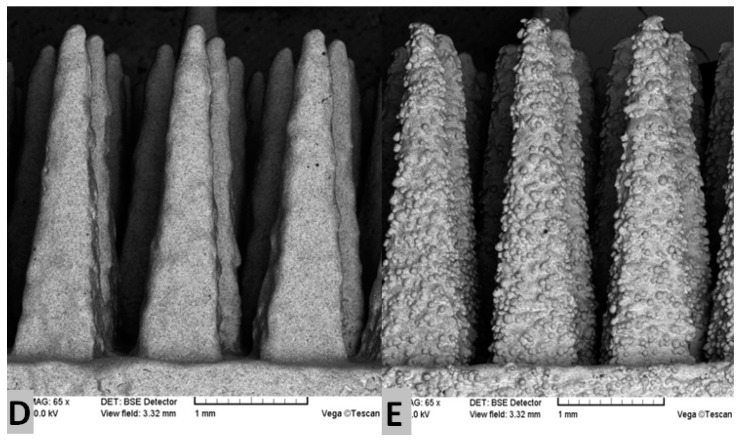
We present here-designed, manufactured, and tested by our research team-the Ti-alloy prototype of the multispiked connecting scaffold (MSC-Scaffold) interfacing the components of resurfacing arthroplasty (RA) endoprostheses with bone. The spikes of the MSC-Scaffold prototype mimic the interdigitations of the articular subchondral bone, which is the natural biostructure interfacing the articular cartilage with the periarticular trabecular bone. To enhance the osteoinduction/osteointegration potential of the MSC-Scaffold, the attempts to modify its bone contacting surfaces by the process of electrochemical cathodic deposition of Ca-P was performed with further immersion of the MSC-Scaffold prototypes in SBF in order to transform the amorphous calcium-phosphate coating in hydroxyapatite-like (HA-like) coating. The pilot experimental study of biointegration of unmodified and Ca-P surface-modified MSC-Scaffold prototypes was conducted in an animal model (swine) and in osteoblast cell culture. On the basis of a microscope-histological method the biointegration was proven by the presence of trabeculae in the interspike spaces of the MSC-Scaffold prototype on longitudinal and cross-sections of bone-implant specimens. The percentage of trabeculae in the area between the spikes of specimen containing Ca-P surface modified scaffold prototype observed in microCT reconstructions of the explanted joints was visibly higher than in the case of unmodified MSC-Scaffold prototypes. Significantly higher Alkaline Phosphatase (ALP) activity and the cellular proliferation in the case of Ca-P-modified MSC-Scaffold pre-prototypes, in comparison with unmodified pre-prototypes, was found in osteoblast cell cultures. The obtained results of experimental implantation in an animal model and osteoblast cell culture evaluations of Ca-P surface-modified and non-modified biomimetic MSC-Scaffold prototypes for biomimetic entirely-cementless RA endoprostheses indicate the enhancement of the osteoinduction/osteointegration potential by the Ca-P surface modification of the Ti-alloy MSC-Scaffold prototype. Planned further research on the prototype of this biomimetic MSC-Scaffold for a new generation of RA endoprostheses is also given.

Keywords: animal model evaluation; bone-implant biomimetic prototype interface; multispiked connecting scaffold (MSC-Scaffold) Ti-alloy prototype; osteoblast cell culture evaluation; osteoinduction and osteointegration potential; resurfacing arthroplasty RA endoprostheses.


Conflict of interest statement

The authors declare no conflict of interest.

Figures

Figure 1
Figure 1
(A,B) Prototype of the biomimetic entirely-cementless total resurfacing hip arthroplasty (TRHA) endoprothesis with the multispiked connecting scaffold (MSC-Scaffold) manufactured with Selective Laser Technology (SLM); (C) the exemplary pre-prototype of the MSC-Scaffold directly after the SLM manufacturing process; (D) the SEM photograph showing numerous micro residues in the form of unmelted powder particles and sphere-like shapes on the multi-spiked surface of the scaffold; and (E) the spikes of the MSC-Scaffold after the manual post-production blasting treatment of its multi-spiked surface with use of an experimentally-customized abrasive mixture.
Figure 1
Figure 1
(A,B) Prototype of the biomimetic entirely-cementless total resurfacing hip arthroplasty (TRHA) endoprothesis with the multispiked connecting scaffold (MSC-Scaffold) manufactured with Selective Laser Technology (SLM); (C) the exemplary pre-prototype of the MSC-Scaffold directly after the SLM manufacturing process; (D) the SEM photograph showing numerous micro residues in the form of unmelted powder particles and sphere-like shapes on the multi-spiked surface of the scaffold; and (E) the spikes of the MSC-Scaffold after the manual post-production blasting treatment of its multi-spiked surface with use of an experimentally-customized abrasive mixture.
Figure 2
Figure 2
(A,B) SEM images of the MSC-Scaffold pre-prototypes subjected to electrochemical modification at a current density 5 mA/cm2 followed by 24 h immersion in SBF, the arrows show the plate-like shaped hydroxyapatite-like crystals on the lateral at the lateral surface of the MSC-Scaffold’s spikes (A), as well as at the base of the scaffold near the spikes’ edges (B); (C,D) the exemplary region on the spikes’ lateral surface and the photograph showing the same region with mapping performed using a specialized software analyzer enclosed in an EDS system; the red color corresponds to atoms of calcium, and green and light-green, to phosphorus atoms; and (E) the exemplary EDS spectrogram dealing with a region on the spikes’ lateral surface, and the corresponding chemical composition showing that the Ca/P ratio is equal to 1.59.
Figure 3
Figure 3
(A) The specimen with the implanted two pre-prototypes of the MSC-Scaffold explanted surgically at eight weeks after the implantation; the lateral (B) and anteroposterior (C) 2D digital X-ray radiograms of the explanted swine knee joint.
Figure 4
Figure 4
View of the window showing 3D micro-CT reconstruction of bone-implant specimen with sections in three dimensions and the exemplary 3D view of the MSC-Scaffold with unmodified spikes’ surface with distinction of the given elements, such as implant (black), trabeculae (T), and bone marrow (BM).
Figure 5
Figure 5
(A) The bone-implant specimens with the unmodified (1) surface and the Ca-P-modified (2) surface of the MSC-Scaffold pre-prototypes cut from the harvested knee joint; (B) the exemplary thin sections and the exemplary histopathological documentation of bone-implant specimens made on these bone-implant thin sections in longitudinal (C) and crosswise directions (D) to the axes of spikes (presented near to particular thin sections of unmodified surface and Ca-P-modified).
Figure 6
Figure 6
Fluorescent micrographs of five-day osteoblast culture after Hoechst 33342 and propidium iodide staining: (A) the unmodified and (B) the Ca-P-modified PSc350 pre-prototypes of the MSC-Scaffold (10× magnification); and (C) the PSc350 pre-prototype of the MSC-Scaffold (20× magnification); the dashed line indicates the contour of a spike base of the tested MSC-Scaffold pre-prototypes as seen from spike top.
Figure 7
Figure 7
ALP enzyme activity in time of incubation in human bone cell line culture, and depending on examined MSC-Scaffold pre-prototype construction variant: PSc200 with unmodified surface, PSc350 with unmodified surface, and with Ca-P-modified surface.

Similar articles

Cited by

References

    1. Sershon R., Balkissoon R., Della Valle C.J. Current indications for hip resurfacing arthroplasty in 2016. Curr. Rev. Musculoskelet. Med. 2016;9:84–92. doi: 10.1007/s12178-016-9324-0. - DOI - PMC - PubMed
    1. Baker R., Whitehouse M., Kilshaw M., Pabbruwe M., Spencer R., Blom A., Bannister G. Maximum temperatures of 89 °C recorded during the mechanical preparation of 35 femoral heads for resurfacing. Acta Orthop. 2011;82:669–673. doi: 10.3109/17453674.2011.636681. - DOI - PMC - PubMed
    1. Asaad A., Hart A., Khoo M.M., Ilo K., Schaller G., Black J.D., Muirhead-Allwood S. Frequent femoral neck osteolysis with Birmingham mid-head resection resurfacing arthroplasty in young patients. Clin. Orthop. Relat. Res. 2015;473:3770–3778. doi: 10.1007/s11999-015-4348-0. - DOI - PMC - PubMed
    1. Cadossi M., Tedesco G., Sambri A., Mazzotti A., Giannini S. Hip resurfacing implants. Orthopedics. 2015;38:504–509. doi: 10.3928/01477447-20150804-07. - DOI - PubMed
    1. Aulakh T.S., Jayasekera N., Singh R., Patel A., Roulahamin N., Kuiper J.H., Richardson J.B. Efficacy of hip resurfacing arthroplasty: 6 Year results from an international multisurgeon prospective cohort study. Acta Orthop. Belg. 2015;81:197–208. - PubMed

LinkOut - more resources