Review
. 2016 Sep 27;9(10):802.
doi: 10.3390/ma9100802.

3D Bioprinting Technologies for Hard Tissue and Organ Engineering

Affiliations
Review

3D Bioprinting Technologies for Hard Tissue and Organ Engineering

Xiaohong Wang et al. Materials (Basel). .

Erratum in

Abstract

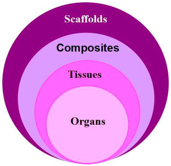
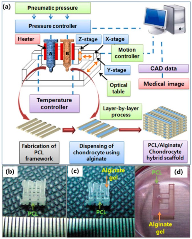
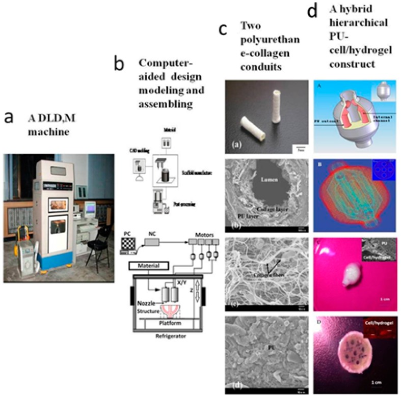
Hard tissues and organs, including the bones, teeth and cartilage, are the most extensively exploited and rapidly developed areas in regenerative medicine field. One prominent character of hard tissues and organs is that their extracellular matrices mineralize to withstand weight and pressure. Over the last two decades, a wide variety of 3D printing technologies have been adapted to hard tissue and organ engineering. These 3D printing technologies have been defined as 3D bioprinting. Especially for hard organ regeneration, a series of new theories, strategies and protocols have been proposed. Some of the technologies have been applied in medical therapies with some successes. Each of the technologies has pros and cons in hard tissue and organ engineering. In this review, we summarize the advantages and disadvantages of the historical available innovative 3D bioprinting technologies for used as special tools for hard tissue and organ engineering.

Keywords: bones; cartilage; composite materials; hard tissues and organs; mechanical properties; teeth.


Conflict of interest statement

The authors declare no conflict of interest. The founding sponsors had no role in the design of the study; in the collection, analyses, or interpretation of data; in the writing of the manuscript, and in the decision to publish the results.

Figures

Figure 1
Figure 1
Applications of 3D printing technologies in regenerative medicine: the produced 3D objects can be porous scaffolds, cell/biomaterials composites, homogeneous tissues, or multiple tissues contained organs.
Figure 2
Figure 2
Working principles of three main groups of bioprinting technologies for tissue and organ engineering: (a) laser-based bioprinting; (b) inkjet-based bioprinting; and (c) extrusion-based bioprinting [22].
Figure 3
Figure 3
(a) 3D printing schematic using an inkjet printing system; and (b) 3D printed calcium phosphate (CaP) sintered structures fabricated at Washington State University using a 3D printer (ProMetal®, ExOne LLC, Irwin, PA, USA) [42].
Figure 4
Figure 4
Schematic of stereolithographic (SLA) printing technique; and (AD) exemplary tissue engineering scaffold composed of poly(d-l lactic acid) (PDLLA) that showcases the resolution and detail of SLA [47]: (A) photograph; (B) micro computed tomography (mCT); and (C,D) scanning electron microscope (SEM). Scale bar is 500 mm.
Figure 5
Figure 5
(a) Schematics of the fabrication process of cell-printed 3D polycaprolactone (PCL)–alginate gel hybrid scaffold using a multihead deposition system; Photo-images of: (b) fabricated porous 3D PCL scaffold; (c) chondrocyte-printed 3D PCL–alginate gel hybrid scaffold for in vivo experiments; and (d) simplified 2D hybrid scaffold for in vitro experiments [57].
Figure 6
Figure 6
A double-nozzle low-temperature (DLDM) technology developed at Tsinghua University, prof. Wang’ group: (a) the DLDM printer; (b) schematic description of the working processes of the two nozzles; (c) a tubular polyurethane-collagen conduit made by the DLDM system; and (d) an elliptical hybrid hierarchical polyurethane and cell/hydrogel construct made by the DLDM system [12].

References

    1. Yu W.Y. The structure, development and health of the teeth. Bull. Biol. 1982;8:36–39.
    1. Eckstein F., Reiser M., Englmeier K.H., Putz R. In vivo morphometry and functional analysis of human articular cartilage with quantitative magnetic resonance imaging—From image to data, from data to theory. Anat. Embryol. 2001;203:147–173. doi: 10.1007/s004290000154. - DOI - PubMed
    1. Wang X.H., Ma J.B., Wang Y.N., He B.L. Progress in the research of bone substitutes. J. Biomed. Eng. 2001;18:647–652. - PubMed
    1. Wang X., Ma J., Feng Q., Cui F.Z. Skeletal repair in rabbits with calcium phosphate cements incorporated phosphorylated chitin. Biomaterials. 2002;23:4591–4600. doi: 10.1016/S0142-9612(02)00205-3. - DOI - PubMed
    1. Wang X., Ma J., Wang Y., He B. Bone repair in radii and tibias of rabbits with phosphorylated chitosan reinforced calcium phosphate cements. Biomaterials. 2002;23:4167–4176. doi: 10.1016/S0142-9612(02)00153-9. - DOI - PubMed