Review
. 2016 Dec 8;9(12):994.
doi: 10.3390/ma9120994.

Fc-Binding Ligands of Immunoglobulin G: An Overview of High Affinity Proteins and Peptides

Affiliations
Review

Fc-Binding Ligands of Immunoglobulin G: An Overview of High Affinity Proteins and Peptides

Weonu Choe et al. Materials (Basel). .

Abstract

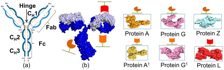
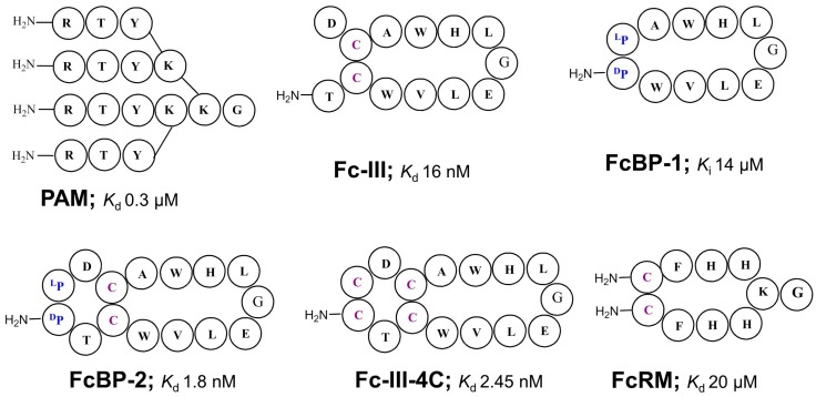
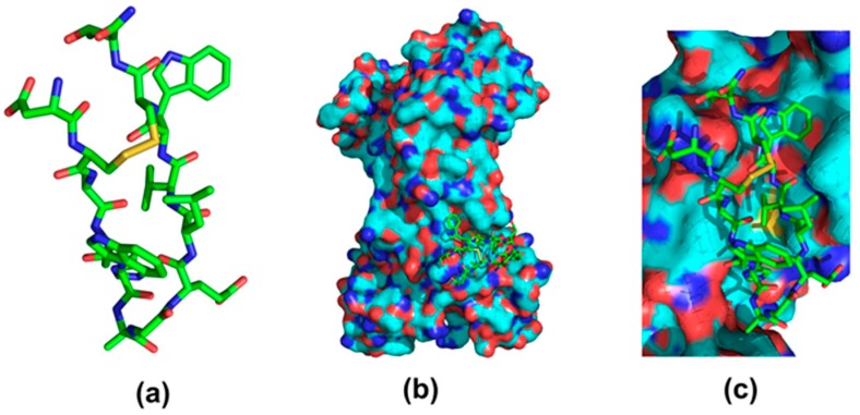
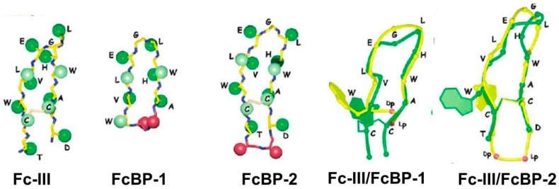
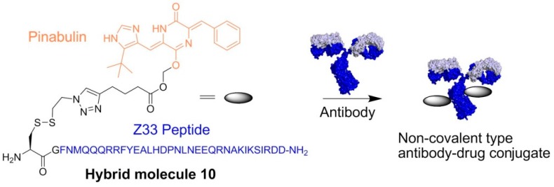
The rapidly increasing application of antibodies has inspired the development of several novel methods to isolate and target antibodies using smart biomaterials that mimic the binding of Fc-receptors to antibodies. The Fc-binding domain of antibodies is the primary binding site for e.g., effector proteins and secondary antibodies, whereas antigens bind to the Fab region. Protein A, G, and L, surface proteins expressed by pathogenic bacteria, are well known to bind immunoglobulin and have been widely exploited in antibody purification strategies. Several difficulties are encountered when bacterial proteins are used in antibody research and application. One of the major obstacles hampering the use of bacterial proteins is sample contamination with trace amounts of these proteins, which can invoke an immune response in the host. Many research groups actively develop synthetic ligands that are able to selectively and strongly bind to antibodies. Among the reported ligands, peptides that bind to the Fc-domain of antibodies are attractive tools in antibody research. Besides their use as high affinity ligands in antibody purification chromatography, Fc-binding peptides are applied e.g., to localize antibodies on nanomaterials and to increase the half-life of proteins in serum. In this review, recent developments of Fc-binding peptides are presented and their binding characteristics and diverse applications are discussed.

Keywords: Fc-binding peptide; Protein A; Protein A mimics; Protein G; Protein L; affinity column chromatography; antibody; targeted drug delivery.


Conflict of interest statement

The authors declare no conflict of interest.

Figures

Figure 1
Figure 1
Illustration of an antibody and the most common bacterial proteins used. (a) Schematic representation of the different parts of an antibody; (b) pymol-generated crystal structure of IgG (pdb id: 1IGT), Protein A (pdb id: 1FC2), Protein G (pdb id: 1FCC), Protein Z (pdb id: 1Q2N), Protein A1 (pdb id: 1DEE), Protein G1 (pdb id: 1QKZ), and Protein L (pdb id: 1HEZ).
Figure 2
Figure 2
Structures of branched- and cyclic peptide-based high affinity ligands that bind to IgGs through interaction with the Fc-domain. The reported affinity constants are towards IgG. Elution pH of antibody from affinity ligand column: PAM, 3 or 9; Fc-III, 3.5; Fc-III-4C, 3.5; FcRM, 2.7 [42,45,50,51,54].
Figure 3
Figure 3
Structure of Fc-III binding to the Fc-region of IgG. (a) Fc-III (DCAWHLGELVWCT-NH2); (b) Full view; (c) close view. Pymol generated image from a PDB file (1DN2).
Figure 4
Figure 4
Superimposition of the backbone atoms of the bound structure of Fc-III (green). Adapted from [50], with permission from © 2006 ACS publications.
Figure 5
Figure 5
(a) Illustration of antibody binding on a solid surface using Fc-binding peptide Fc-III; (b) SDS-PAGE analysis of IgGs bound to Fc-III covered magnetic beads. Human IgG1 (Kd = 85 ± 5 nM), mouse IgG3 (Kd > 20 μM), goat IgG3 (Kd not given), and rabbit IgG (Kd = 305 ± 37 nM) were incubated with Fc-III functionalized (+) or unmodified (−) magnetic beads. After washing steps with PBS, the bound IgG were applied to a 10% SDS-polyacrylamide gel. Protein size marker (M) and 1 μg of human IgG (C) are indicated. Adapted from [70], with permission from © 2016 Elsevier.
Figure 6
Figure 6
Illustration of the complexation of anti-Her2 antibody on a protein nanocage (ferritin) surface using the Fc-binding peptide Fc-III. (a) Ferritin monomer; (b) genetically engineered ferritin fusion monomer with Fc-III inserted in one of the loops; (c) self-assembled ferritin nanocage with 24 surface exposed Fc-III; (d) non-covalent binding of anti-Her2 antibody to the Fc-III functionalized protein nanocage; (e) covalent fluorescent labeling of the protein nanocage.
Figure 7
Figure 7
Fluorescent microscopic images of SKBR3 breast cancer cells (AC) and MCF10A breast cells (DF) treated with Cy3-trastuzumab/fFcBP-Pf_Fn complexes. Trastuzumab and FcBP-Pf_Fn were labeled with Cy3 (Cy3-trastuzumab) and fluorescein (fFcBP-Pf_Fn), respectively. Cy3-trastuzumab was simply mixed with fFcBP-Pf_Fn to form Cy3-trastuzumab/fFcBP-Pf_Fn complexes and subsequently incubated with SKBR3 breast cancer cells and MCF10A breast cells in the presence of excess amounts of rabbit serum. Cy3-trastuzumab and fFcBP-Pf_Fn are visualized as orange and green, respectively, by fluorescence microscopy. (For interpretation of the references to color in this figure legend, the reader is referred to the web version of this article.) Adapted from [71], with permission from © 2012 Elsevier.
Figure 8
Figure 8
(a) Illustration of fluorescent labeled antibody delivery into live cells using the Fc-domain binding peptide (FcBP) Fc-III conjugated to the cell penetrating peptide Tat in addition to the green fluorescent protein; (b) Delivery of human IgG into live cells using the Fc-III/eGFP/Tat constructs. Cells were treated with Cy3 labelled human IgG (Cy3hIgG) in the presence of the Fc-III/eGFP/Tat or Fc-III/Tat. Cy3 (red): the fluorescence images of Cy3-hIgG (Ex/Em = 555/605 nm); EGFP: the fluorescence of eGFP the fluorescence (green) of eGFP (Ex/Em = 490/525 nm); Nucleus (blue) was stained with DAPI (Ex/Em = 350/455 nm); Cy3/eGFP: superimposition of Cy3-hIgG and FcBP-eGFP-Tat fluorescence images; Cy3/eGFP/DAPI: superimposition of Cy3-hIgG, FcBP-eGFP-Tat and DAPI fluorescence images. (A) HeLa cells treated with Cy3-hIgG and FcBP-eGFP-Tat; (B) HeLa cells treated with Cy3-hIgG and FcBP-Tat; (C) 3T3-L1 cells treated with Cy3-hIgG and FcBP-eGFP-Tat; (D) 3T3-L1 cells treated with Cy3-hIgG and FcBP-Tat. Adapted from [73], with permission from © 2015 Springer.
Figure 9
Figure 9
(a) Schematic of the co-administration scheme. In this experiment, human FcRn Tg mice were not pre-dosed with exogenous hIgG1. Instead mKate-IgGBP and hIgG1 were pre-mixed in a 1:1 molar ratio and co-injected via the tail vein; (b) clearance of mKate-IgGBP in hFcRn Tg mice dosed alone (blue triangles) or co-dosed at a 1:1 molar mixture with hIgG1 (yellow triangles). The mKate-IgGBP remaining (%) was calculated by normalizing the fluorescent emission at all time points to the maximum value observed in the first bleed 5 min after protein injection; (c) clearance of labeled human IgG1 in hFcRn Tg mice dosed as a single agent via the tail vein (blue triangles) compared to the clearance of labeled hIgG1 co-administered as a 1:1 molar mixture with mKate-IgGBP was measured to determine if bound mKate-IgGBP alters the eliminate profile of hIgG1 (red squares). The hIgG1 remaining (%) was calculated by normalizing the fluorescent emission at all time points to the maximum value observed in the first bleed 5 min after protein injection. Dashed lines in each panel represent the data fit to a 2-compartment PK model in Prism and the β-phase half-life shown in the figure was calculated as described in the Methods section. The data shown in each panel are the mean (n = 3 bleeds per time point) and error bars indicate standard deviation. Adapted from [74].
Scheme 1
Scheme 1
Preparation of non-covalent antibody—drug conjugate using Pinabulin (cytotoxic agent) and Fc-binding peptide Z33.
Figure 10
Figure 10
Cytotoxicity of NC-ADC, complex of hybrid 10 and Herceptin, against (A) SKBR-3 cells; (B) MCF-7 cells; and (C) SKBR-3HR cells; (D) cytotoxicity of noncovalent ADC, complex of hybrid 10 and 6E1 against A375 cell. n.s.: not significant, * p < 0.05, ** p < 0.01, *** p < 0.005. Data (n = 3) are shown as means ± SD. Adapted from [75], with permission from © 2016 American Chemical Society.

Similar articles

Cited by

References

    1. Steinitz M. Three decades of human monoclonal antibodies: Past, present and future developments. Hum. Antibodies. 2009;18:1–10. - PubMed
    1. Leavy O. Therapeutic antibodies: Past, present and future. Nat. Rev. Immunol. 2010;10:297. doi: 10.1038/nri2763. - DOI - PubMed
    1. Matera M.G., Page C., Rogliani P., Calzetta L., Cazzola M. Therapeutic monoclonal antibodies for the treatment of chronic obstructive pulmonary disease. Drugs. 2016;76:1257–1270. doi: 10.1007/s40265-016-0625-9. - DOI - PubMed
    1. Mould D.R., Meibohm B. Drug development of therapeutic monoclonal antibodies. BioDrugs. 2016;30:275–293. doi: 10.1007/s40259-016-0181-6. - DOI - PubMed
    1. Lambour J., Naranjo-Gomez M., Piechaczyk M., Pelegrin M. Converting monoclonal antibody-based immunotherapies from passive to active: Bringing immune complexes into play. Emerg. Microbes Infect. 2016;5:e92. doi: 10.1038/emi.2016.97. - DOI - PMC - PubMed

LinkOut - more resources