

Acute uveal effusion during phacoemulsification with preoperative central serous chorioretinopathy: a case report
- PMID: 28774289
- PMCID: PMC5543589
- DOI: 10.1186/s12886-017-0530-3
Acute uveal effusion during phacoemulsification with preoperative central serous chorioretinopathy: a case report
Abstract
Background: We report a case of acute uveal effusion during phacoemulsification in an eye with preoperative chronic central serous chorioretinopathy (CSC).
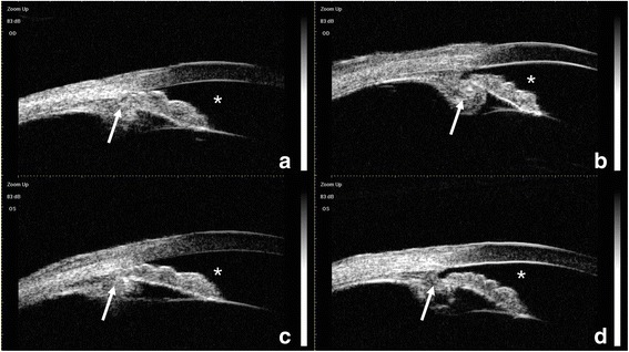
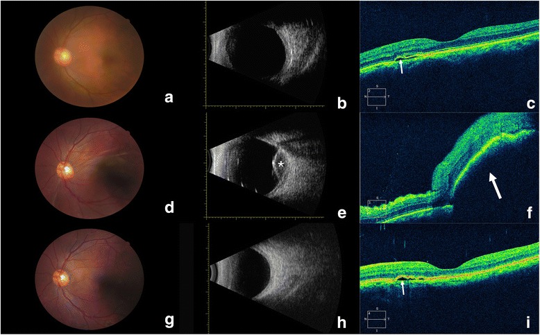
Case presentation: A 55-year-old man with a history of chronic CSC for >18 months presented with bilateral opaque lenses. A preoperative ophthalmic examination showed suspected lenticonus and risky anatomical features, including a thick ciliary body, and anterior rotation of the ciliary process and iris root in both eyes. Optical coherence tomography (OCT) detected CSC in the left eye, but the results of fundus photography and B-scan ultrasonography were unremarkable. The anterior chamber flattened during phacoemulsification. Anterior vitrectomy was quickly performed for suspected infusion misdirection syndrome, and was followed by uneventful surgery. On postoperative day 1, fundus photography, type B ultrasound, and OCT revealed uveal exudation in the macula of the left eye. On postoperative day 50, the patient's visual acuity recovered to 20/32, and fundus photography, ultrasonography, and OCT revealed complete resolution of the uveal effusion.
Conclusions: This case suggests an association between preoperative CSC and uveal effusion during surgery, because choroidal hyperperfusion and hyperpermeability were present in the patient's CSC-affected eyes.
Keywords: Case report; Central serous chorioretinopathy; Choroidal hyperperfusion; Hyperpermeability; Infusion misdirection syndrome; Suprachoroidal hemorrhage; Uveal effusion.
Conflict of interest statement
Ethics approval and consent to participate
All procedures in this study that involved human participants were performed in accordance with the ethical standards of the institutional and/or national research committee and with the 1964 Declaration of Helsinki and its later amendments or comparable ethical standards. The Ethics Committee of the Eye and Ear, Nose, and Throat Hospital, Fudan University, approved the study.
Consent for publication
The patient provided written consent to publish this case report.
Competing interests
The authors declare that they have no competing interests.
Publisher’s Note
Springer Nature remains neutral with regard to jurisdictional claims in published maps and institutional affiliations.
Figures
References
Publication types
MeSH terms
LinkOut - more resources
Full Text Sources
Other Literature Sources
Medical
