Review
. 2017 Sep:69:158-171.
doi: 10.1016/j.semcdb.2017.07.043. Epub 2017 Aug 1.

Regulation of Wnt signaling by protocadherins

Affiliations
Review

Regulation of Wnt signaling by protocadherins

Kar Men Mah et al. Semin Cell Dev Biol. 2017 Sep.

Abstract

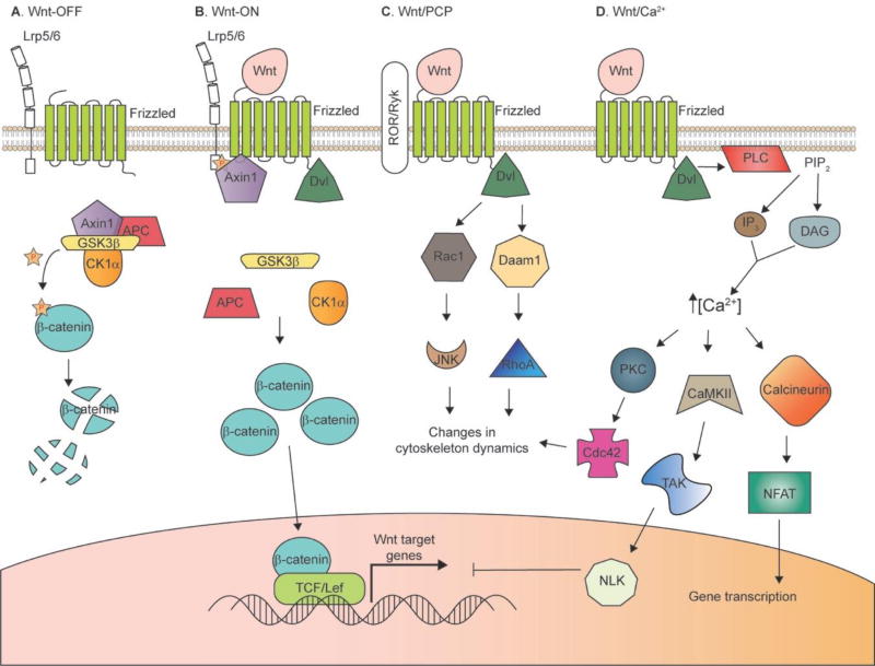
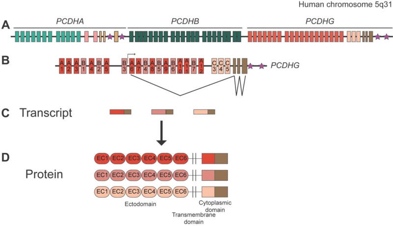
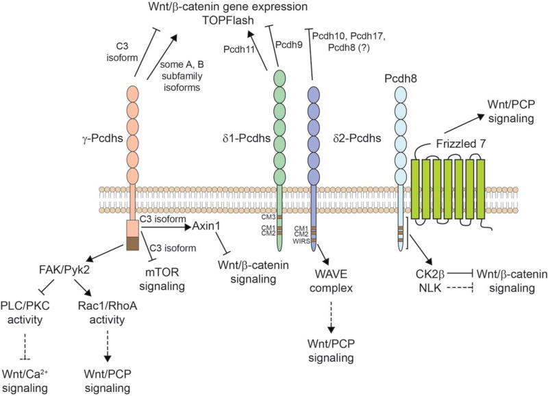
The ∼70 protocadherins comprise the largest group within the cadherin superfamily. Their diversity, the complexity of the mechanisms through which their genes are regulated, and their many critical functions in nervous system development have engendered a growing interest in elucidating the intracellular signaling pathways through which they act. Recently, multiple protocadherins across several subfamilies have been implicated as modulators of Wnt signaling pathways, and through this as potential tumor suppressors. Here, we review the extant data on the regulation by protocadherins of Wnt signaling pathways and components, and highlight some key unanswered questions that could shape future research.

Keywords: Cancer; Cell adhesion; Epigeneticsextracellular cadherin (EC); Planar cell polarity; Tumor suppressor.


Figures

Figure 1
Figure 1. Wnt signaling pathways
A and B: Canonical (β-catenin-dependent) pathway. In the “Wnt-OFF” state (A), β-catenin levels are kept low in the cytoplasm by the action of the “destruction complex”. GSK3β phosphorylates β-catenin, which targets it for desctruction by the proteasome. Wnt binding to Frizzled and Lrp5/6 co-receptors results in disruption of the destruction complex, allowing β-catenin to accumulate in the cytoplasm. β-catenin can then translocate to the nucleus and promote the activation of Wnt target genes by displacing co-repressors, and recruiting co-activators, of TCF/Lef. C: Wnt/PCP pathway. Binding of Wnt to Frizzled and ROR or Ryk co-receptors leads to recruitment of Dvl, which can act through Rac1 or Daam1 to initate changes in cytoskeletal dynamics important for cell orientation and movement. D: Wnt/Ca2+ pathway. Wnt binding and recruitment of Dvl leads to activation of PLC, which cleaves PIP2 to generate IP3 and DAG. This leads to release of Ca2+ from intracellular stores, and downstream signaling through a number of Ca2+-dependent kinases and phosphatases.
Figure 2
Figure 2. The protocadherin gene clusters
A: Schematic of the human PCDHA, PCDHB, and PCDHG gene clusters on chromosome 5q31. A very similar structure is observed for the mouse clusters at chromosome 18. B: The exon structure of the PCDHG cluster is expanded below, with an example of the transcription initiation and splicing pattern (for A6, in this instance). C: Schematic of the PCDHG spliced transcripts generated by the cluster; each mature transcript consists of one large variable exon and the three small constant exons. D: Protein structure of the γ-Pcdhs (α-Pcdhs are identical in structure; β-Pcdhs lack any constant domain). Six extracellular cadherin (EC) repeats, a transmembrane domain, and a variable cytoplasmic domain are encoded by each variable exon; the constant exons encode a 125 amino acid C-terminal domain. Stars indicate the sites of “cluster control regions”, enhancers required for normal expression patterns of the Pcdh clusters.
Figure 3
Figure 3. Regulation of Wnt pathways by Pcdhs
Summary of results implicating γ-Pcdhs (left) and δ-Pcdhs (right) in the regulation of Wnt signaling, as discussed in the main text. The γ-Pcdh-C3 isoform, and the δ-Pcdhs Pcdh8, 9, 10, and 17 have been reported to suppress Wnt-induced expression of target genes, while some other γ-Pcdh isoforms and the δ1 protein Pcdh11 have been reported to have the opposite effect (top). Some Pcdhs have known cytoplasmic interactors that have been shown to, or that potentially could, impinge upon Wnt signaling pathways (bottom). Pcdh8 has been shown to interact with Frizzeld7 to promote Wnt/PCP signaling (far right). Long lines with a short perpendicular line indicate inhibition, while arrows indicate activation. Dashed lines indicate possible signaling connections, based on the literature, that remain to be demonstrated directly.

Similar articles

Cited by

References

    1. Peek SL, Mah KM, Weiner JA. Regulation of neural circuit formation by protocadherins. Cellular and Molecular Life Sciences. 2017:1–25. - PMC - PubMed
    1. Keeler AB, Molumby MJ, Weiner JA. Protocadherins branch out: Multiple roles in dendrite development. Cell Adh Migr. 2015;9(3):214–26. - PMC - PubMed
    1. Angers S, Moon RT. Proximal events in Wnt signal transduction. Nature reviews Molecular cell biology. 2009;10(7):468–477. - PubMed
    1. Cadigan KM, Peifer M. Wnt signaling from development to disease: insights from model systems. Cold Spring Harbor perspectives in biology. 2009;1(2):a002881. - PMC - PubMed
    1. Clevers H, Nusse R. Wnt/β-catenin signaling and disease. Cell. 2012;149(6):1192–1205. - PubMed

Publication types

LinkOut - more resources