

Advancing imaging technologies for patients with spinal pain: with a focus on whiplash injury
- PMID: 28774580
- PMCID: PMC6874915
- DOI: 10.1016/j.spinee.2017.06.015
Advancing imaging technologies for patients with spinal pain: with a focus on whiplash injury
Abstract
Background context: Radiological observations of soft-tissue changes that may relate to clinical symptoms in patients with traumatic and non-traumatic spinal disorders are highly controversial. Studies are often of poor quality and findings are inconsistent. A plethora of evidence suggests some pathoanatomical findings from traditional imaging applications are common in asymptomatic participants across the life span, which further questions the diagnostic, prognostic, and theranostic value of traditional imaging. Although we do not dispute the limited evidence for the clinical importance of most imaging findings, we contend that the disparate findings across studies may in part be due to limitations in the approaches used in assessment and analysis of imaging findings.
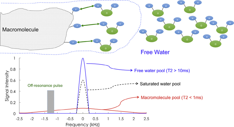
Purpose: This clinical commentary aimed to (1) briefly detail available imaging guidelines, (2) detail research-based evidence around the clinical use of findings from advanced, but available, imaging applications (eg, fat and water magnetic resonance imaging and magnetization transfer imaging), and (3) introduce how evolving imaging technologies may improve our mechanistic understanding of pain and disability, leading to improved treatments and outcomes.
Study design/setting: A non-systematic review of the literature is carried out.
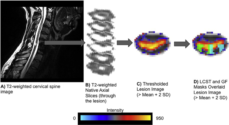
Methods: A narrative summary (including studies from the authors' own work in whiplash injuries) of the available literature is provided.
Results: An emerging body of evidence suggests that the combination of existing imaging sequences or the use of developing imaging technologies in tandem with a good clinical assessment of modifiable risk factors may provide important diagnostic information toward the exploration and development of more informed and effective treatment options for some patients with traumatic neck pain.
Conclusions: Advancing imaging technologies may help to explain the seemingly disconnected spectrum of biopsychosocial signs and symptoms of traumatic neck pain.
Keywords: Biopsychosocial; Imaging; Low back pain; MRI; Neck pain; Whiplash.
Copyright © 2017 Elsevier Inc. All rights reserved.
Figures
References
-
- Manchikanti L, Singh V, Falco FJ, Benyamin RM, Hirsch JA. Epidemiology of low back pain in adults. Neuromodulation 2014;17(Suppl. 2):3–10. - PubMed
-
- Hoy D, Brooks P, Blyth F, Buchbinder R. The Epidemiology of low back pain. Best Pract Res Clin Rheumatol 2010;24:769–81. - PubMed
-
- Hoy D, March L, Brooks P, et al. The global burden of low back pain: estimates from the Global Burden of Disease 2010 study. Ann Rheum Dis 2014;73:968–74. - PubMed
Publication types
MeSH terms
Grants and funding
LinkOut - more resources
Full Text Sources
Other Literature Sources
Medical
