. 2017 Nov 1;313(5):G386-G398.
doi: 10.1152/ajpgi.00215.2017. Epub 2017 Aug 3.

Enteric serotonin and oxytocin: endogenous regulation of severity in a murine model of necrotizing enterocolitis

Affiliations

Enteric serotonin and oxytocin: endogenous regulation of severity in a murine model of necrotizing enterocolitis

Kara Gross Margolis et al. Am J Physiol Gastrointest Liver Physiol. .

Erratum in

  • Corrigendum.
    [No authors listed] [No authors listed] Am J Physiol Gastrointest Liver Physiol. 2018 Mar 1;314(3):G459. doi: 10.1152/ajpgi.zh3-7428-corr.2018. Am J Physiol Gastrointest Liver Physiol. 2018. PMID: 29516747 Free PMC article. No abstract available.

Abstract

Necrotizing enterocolitis (NEC), a gastrointestinal inflammatory disease of unknown etiology that may also affect the liver, causes a great deal of morbidity and mortality in premature infants. We tested the hypothesis that signaling molecules, which are endogenous to the bowel, regulate the severity of intestinal and hepatic damage in an established murine NEC model. Specifically, we postulated that mucosal serotonin (5-HT), which is proinflammatory, would exacerbate experimental NEC and that oxytocin (OT), which is present in enteric neurons and is anti-inflammatory, would oppose it. Genetic deletion of the 5-HT transporter (SERT), which increases and prolongs effects of 5-HT, was found to increase the severity of systemic manifestations, intestinal inflammation, and associated hepatotoxicity of experimental NEC. In contrast, genetic deletion of tryptophan hydroxylase 1 (TPH1), which is responsible for 5-HT biosynthesis in enterochromaffin (EC) cells of the intestinal mucosa, and TPH inhibition with LP-920540 both decrease the severity of experimental NEC in the small intestine and liver. These observations suggest that 5-HT from EC cells helps to drive the inflammatory damage to the gut and liver that occurs in the murine NEC model. Administration of OT decreased, while the OT receptor antagonist atosiban exacerbated, the intestinal inflammation of experimental NEC. Data from the current investigation are consistent with the tested hypotheses-that the enteric signaling molecules, 5-HT (positively) and OT (negatively) regulate severity of inflammation in a mouse model of NEC. Moreover, we suggest that mucosally restricted inhibition of 5-HT biosynthesis and/or administration of OT may be useful in the treatment of NEC.NEW & NOTEWORTHY Serotonin (5-HT) and oxytocin reciprocally regulate the severity of intestinal inflammation and hepatotoxicity in a murine model of necrotizing enterocolitis (NEC). Selective depletion of mucosal 5-HT through genetic deletion or inhibition of tryptophan hydroxylase-1 ameliorates, while deletion of the 5-HT uptake transporter, which increases 5-HT availability, exacerbates the severity of NEC. In contrast, oxytocin reduces, while the oxytocin receptor antagonist atosiban enhances, NEC severity. Peripheral tryptophan hydroxylase inhibition may be useful in treatment of NEC.

Keywords: inflammation; intestine; necrotizing enterocolitis; oxytocin; serotonin.


Figures

Fig. 1.
Fig. 1.
Experimental necrotizing enterocolitis (NEC) leads to weight loss, mortality, and increased transcription of proinflammatory cytokines in intestine and liver. A: control mice gained weight steadily during the 4-day experimental period. The plot of the %weight change as a function of time does not differ significantly from linearity, the slope (m) of the line is positive and deviates significantly from zero (P < 0.001; n = 3). B: experimental mice lost weight steadily during the test period. The plot of the %weight change as a function of time does not differ significantly from linearity, the slope of the line is negative and deviates significantly from zero (P < 0.001; n = 15). The slope of the line in A is significantly different from that in B. C: mean %weight change of surviving control mice at the end of the 4-day test period is significantly greater than that of surviving experimental animals (P < 0.001). D: Kaplan-Meier plots of the survival of control and experimental mice during the course of the 4-day test period. Survival was 100% for control mice and 28.5% for experimental animals. The survival curves are significantly different (Mantel-Cox log-rank test; P < 0.01). E: small intestines of the mice with NEC had multiple strictures (arrows) in the small intestine. The bowel was dilated and gas-filled immediately proximal to the strictures. Strictures were most common just proximal to the cecum. Transcripts encoding proinflammatory cytokines were measured and compared in the distal small intestines of control and experimental mice (NEC). Significant increases were found in the abundance of transcripts encoding: IL-6 (F), IL-1β (G), TNF-α (H), and IL-18 (I). Transcripts encoding proinflammatory cytokines were measured and compared in the livers of NEC mice. Significant increases were found in the abundance of transcripts encoding IL-1β (J) and TNF-α (K).
Fig. 2.
Fig. 2.
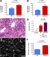
Experimental NEC leads to hepatic inflammation, lipid accumulation, and apoptosis of hepatocytes. A: liver of a mouse subjected to the control protocol and stained with hematoxylin and eosin (H&E). The architecture of the liver, hepatocytes lining sinusoids, portal area, and central vein are all normal. The hepatocytes are not vacuolated (see also inset). Bar = 100 µm. B: liver of a mouse subjected to the NEC protocol and stained with H&E. An infiltrate of inflammatory cells surrounds a bile ductule in a portal area (see white arrow). Hepatocytes appear vacuolated (see also inset). Bar = 50 µm. C: histological score of inflammation is compared in sections of the livers of mice subjected to the control and NEC protocols. Significant inflammation was evident in the livers of mice exposed to NEC but not in those of control animals. D: frozen section of the liver of a mouse subjected to the control protocol and stained with Sudan Black. Very little structure is evident because the tissue lacks accumulated sudanophilic lipid. Bar = 50 µm. E: frozen section of the liver of a mouse subjected to the NEC protocol and stained with Sudan Black. Hepatocytes of the hepatic parenchyma are intensely sudanophilic, confirming that the vacuolization evident in H&E section is due to the presence of lipid droplets in the hepatocyte cytoplasm. Bar = 50 µm. F: densitometric quantitation of the Sudan Black staining of the livers of mice subjected to the control and NEC protocols. Hepatocytes of mice subjected to the NEC protocol were significantly more sudanophilic than those of control animals. Note that the scale of the ordinate is logarithmic. G: TUNEL staining of the liver of a mouse subjected to the control protocol reveals little or no apoptosis. Bar = 100 µm. H: TUNEL staining of the liver of a mouse subjected to the NEC protocol reveals an area where many hepatocytes are undergoing apoptosis. Bar = 100 µm. H, inset: nuclei of hepatocytes are filled with TUNEL reaction product. Small mononuclear cells surround the apoptotic hepatocytes. I: quantitation of the area of hepatic parenchyma containing apoptotic hepatocytes in the livers of mice subjected to the control and NEC protocols. Hepatocytes of mice subjected to the NEC protocol undergo significantly more apoptosis than those of control animals. Note that the ordinate is logarithmic.
Fig. 3.
Fig. 3.
Intestinal inflammation is more severe in SERTKO than in WT mice after exposure to the NEC protocol. A: percent change in weight as a function of time is compared in WT and SERTKO mice subjected to the control and NEC protocols. Both WT (n = 10) and SERTKO (n = 10) mice gain weight during the control protocol; the difference between the two groups is not significant. In contrast, both WT (n = 36) and SERTKO (n = 40) mice lose weight during the NEC protocol; however, SERTKO mice lose significantly more weight than their WT counterparts. Photographs of the abdominal contents of a WT (B) and a SERTKO (C) mouse after exposure for 5 days to the NEC protocol. Loops of bowel are dilated with gas in both; however, the severity of the gaseous dilation is greater in the SERTKO than in the WT animal. Expression of the proinflammatory cytokines normalized to that of GAPDH, IL-18 (D) and IL-6 (E), as well as iNOS (F) are all significantly more elevated in the intestines of SERTKO than in WT mice.
Fig. 4.
Fig. 4.
Hepatic inflammation is more severe in SERTKO than in WT mice after exposure to the NEC protocol. A: transcripts encoding IL-1β are more abundant in the livers of SERTKO than WT mice. B: transcripts encoding TNF-α are more abundant in the livers of SERTKO than WT mice. C: periportal infiltration of inflammatory cells in a SERTKO mouse (compare with Fig. 2B (NEC in a WT mouse)]. Bar = 100 μm. The cytoplasm of hepatocytes appears vacuolated. D: histological score of inflammation is compared in sections of the livers of WT and SERTKO mice subjected to the control and NEC protocols. Significantly higher inflammatory scores were found in the livers of SERTKO mice exposed to the NEC protocol but not in any animals subjected to the control protocol. E: frozen section of the liver of a SERTKO mouse subjected to the NEC protocol and stained with Sudan Black. Hepatocytes of the hepatic parenchyma are intensely sudanophilic [compare with Fig. 2E (NEC in a WT mouse)]. Bar = 100 μm. F: densitometric quantitation of the Sudan Black staining of the livers of WT and SERTKO mice subjected to the NEC protocol. Hepatocytes of SERTKO mice subjected to the NEC protocol were significantly more sudanophilic than those of WT animals.
Fig. 5.
Fig. 5.
Intestinal inflammation and hepatic toxicity are less severe in TPH1KO than in WT mice. A: percent change in weight as a function of time in mice subjected to control protocol (n = 10) is compared with both WT (n = 28) and TPH1KO (n = 28) mice subjected to the NEC protocol. Mice gained weight during the control protocol (compare with Fig. 3A (WT and SERTKO mice subjected to the control protocol). In contrast, both TPH1KO and WT mice lost weight during the NEC protocol; however, WT mice lost significantly more weight than their TPH1KO counterparts. B: transcripts encoding IL-6 are significantly more abundant in the intestines of mice subjected to the NEC than the control protocol; however, the increase in abundance of enteric IL-6 transcripts is significantly greater in WT than in TPH1KO mice. The NEC-induced increase in transcripts encoding IL-6 in the intestines of TPH1KO mice is not significantly different from control. C: transcripts encoding IL-18 are significantly more abundant in mice subjected to the NEC than the control protocol; however, the increase in the abundance of IL-18 transcripts is significantly greater in WT than in TPH1KO mice. D: transcripts encoding TNF-α are significantly more abundant in the livers of mice subjected to the NEC than the control protocol; however, the increase in abundance of TNF-α transcripts is significantly greater in WT than in TPH1KO mice. The NEC-induced increase in transcripts encoding TNF-α in the livers of TPH1KO mice is not significantly different from control. E: transcripts encoding IL-1β are significantly more abundant in the livers of mice subjected to the NEC than the control protocol; however, the increase in abundance of IL-1β transcripts is significantly greater in WT than in TPH1KO mice. F: Sudanophilia is significantly more abundant in the livers of mice subjected to the NEC than the control protocol; however, the degree of sudanophilia is significantly greater in WT than in TPH1KO mice. The NEC-induced increase in sudanophilia in the livers of TPH1KO mice is not significantly different from control. G: apoptosis, detected with the TUNEL reaction is significantly more abundant in the livers of mice subjected to the NEC than the control protocol; however, the frequency of apoptosis is significantly greater in WT than in TPH1KO mice. The NEC-induced increase in apoptosis in the livers of TPH1KO mice is not significantly different from that in the livers of control animals. H: illustrations of the TUNEL reactivity in the livers of NEC-exposed TPH1KO (left) and WT (right) mice (compare with the quantified data in G; see arrow). Note that TPH1KO protects the liver from the effects of the NEC protocol. I: frozen sections of the livers of a TPH1KO (left) and WT mouse (right) subjected to the NEC protocol and stained with Sudan Black. Hepatocytes of the hepatic parenchyma are more intensely sudanophilic in the liver of the WT than in the TPH1KO mouse (compare with the quantified data in F; see arrow). H and I: bar = 100 μm.
Fig. 6.
Fig. 6.
The mucosally restricted TPH inhibitor, LP-920540, decreases the severity of the effects of the NEC protocol in the intestine and liver. A: Kaplan-Meier plots of the survival of WT mice treated with vehicle (n = 21) or LP-920540 (n = 42) after exposure to the NEC protocol. Survival was significantly better in animals treated with 100 (n = 21) or 200 mg/kg (n = 21) LP-902540 than in those treated with vehicle; the difference in survival between the two doses of LP-920540 is not significant. B: percent change in weight as a function of time is compared in WT mice treated with vehicle or LP-920540. Both vehicle- and LP-920540-treated mice lose weight during the NEC protocol; however, vehicle-treated mice lose significantly more weight than their LP-920540-treated counterparts. The 200 mg/kg dose of LP-920540 provides significantly better protection from NEC than 100 mg/kg. C: histological scores of intestinal inflammation in mice subjected to the NEC protocol are compared between vehicle- and LP-920540-treated animals. Inflammation is less severe in LP-920540-treated mice (200 mg/kg provides significantly better protection that 100 mg/kg). D and E: representative H&E-stained sections of the colons of mice subjected to the NEC protocol. Inflammation and tissue damage (vacuolization and villous blunting) are less severe in the colon of a mouse treated with 200 mg/kg LP-920540 (D) than vehicle (E). Transcripts encoding the proinflammatory cytokines IL-18 (F) and IL-6 (G) were measured and compared in the distal small intestines of vehicle- and LP-920540-treated mice undergoing the control and NEC protocols. Abundance of each transcript was greater in vehicle- than in LP-920540-treated animals undergoing the NEC protocol. H: abundance of transcripts encoding TNF-α in the livers of mice undergoing the control protocol is compared with that in vehicle- and LP-920540 (200 mg/kg)-treated mice subjected to the NEC protocol. LP-920540 provides significant protection of the liver from the effects of NEC. I: abundance of transcripts encoding IL-1β in the livers mice undergoing the control protocol is compared with that in vehicle- and LP-920540 (200 mg/kg)-treated mice subjected to the NEC protocol. LP-920540 provides significant protection of the liver from the effects of NEC. J: intensity of sudanophilia in the hepatocytes of the livers of mice undergoing the NEC protocol is significantly greater in vehicle- than in LP-920540-treated mice. K: amount of apoptosis, as measured by TUNEL assay, in the hepatocytes of the livers of mice undergoing the NEC protocol is significantly greater in vehicle- than in LP-920540-treated mice.
Fig. 7.
Fig. 7.
Oxytocin attenuates the severity of the effects of the NEC protocol on mouse intestine. Transcripts encoding proinflammatory molecules related to inflammation are less abundant in the NEC-exposed mouse colon in OT-treated mice (n = 9) than in those receiving vehicle (n = 48). A: gene expression in a focused microarray. The abundance of transcripts in OT-treated mice is plotted logarithmically on the ordinate as a function of the abundance of the same transcripts in vehicle-treated mice plotted logarithmically on the abscissa. The central line shows the best fit of the data, and the parallel lines above and below depict twofold limits. Many genes (green dots below the lower limiting line) are expressed to a lesser extent in OT- than in vehicle-treated mice, but only two genes (red dots above the upper limiting line) are more highly expressed in OT-treated animals. Both of these gene products Ccr4 and Csf3 are anti-inflammatory or immunosuppressive. B–D: transcripts encoding IL-1β (B), IL-6 (C), and TNF-α (D) are significantly more abundant in the intestine of atosiban- (n = 9) than vehicle-treated (n = 48) mice subjected to the NEC protocol. Transcripts encoding IL-1β (E), IL-6 (F), and TNF-α (G) are significantly more abundant in the intestine of vehicle- than OT-treated mice after exposure to the NEC protocol.

Similar articles

Cited by

References

    1. Afrazi A, Branca MF, Sodhi CP, Good M, Yamaguchi Y, Egan CE, Lu P, Jia H, Shaffiey S, Lin J, Ma C, Vincent G, Prindle T Jr, Weyandt S, Neal MD, Ozolek JA, Wiersch J, Tschurtschenthaler M, Shiota C, Gittes GK, Billiar TR, Mollen K, Kaser A, Blumberg R, Hackam DJ. Toll-like receptor 4-mediated endoplasmic reticulum stress in intestinal crypts induces necrotizing enterocolitis. J Biol Chem 289: 9584–9599, 2014. doi:10.1074/jbc.M113.526517. - DOI - PMC - PubMed
    1. Azcarate-Peril MA, Foster DM, Cadenas MB, Stone MR, Jacobi SK, Stauffer SH, Pease A, Gookin JL. Acute necrotizing enterocolitis of preterm piglets is characterized by dysbiosis of ileal mucosa-associated bacteria. Gut Microbes 2: 234–243, 2011. doi:10.4161/gmic.2.4.16332. - DOI - PMC - PubMed
    1. Bertrand PP, Kunze WAA, Furness JB, Bornstein JC. The terminals of myenteric intrinsic primary afferent neurons of the guinea-pig ileum are excited by 5-hydroxytryptamine acting at 5-hydroxytryptamine-3 receptors. Neuroscience 101: 459–469, 2000. doi:10.1016/S0306-4522(00)00363-8. - DOI - PubMed
    1. Bischoff SC, Mailer R, Pabst O, Weier G, Sedlik W, Li Z, Chen JJ, Murphy DL, Gershon MD. Role of serotonin in intestinal inflammation: knockout of serotonin reuptake transporter exacerbates 2,4,6-trinitrobenzene sulfonic acid colitis in mice. Am J Physiol Gastrointest Liver Physiol 296: G685–G695, 2009. doi:10.1152/ajpgi.90685.2008. - DOI - PubMed
    1. Blakely RD, De Felice LJ, Hartzell HC. Molecular physiology of norepinephrine and serotonin transporters. J Exp Biol 196: 263–281, 1994. - PubMed

MeSH terms

Substances

LinkOut - more resources