. 2017 Aug 4;357(6350):503-507.
doi: 10.1126/science.aan2475.

Chemogenetics revealed: DREADD occupancy and activation via converted clozapine

Affiliations

Chemogenetics revealed: DREADD occupancy and activation via converted clozapine

Juan L Gomez et al. Science. .

Abstract

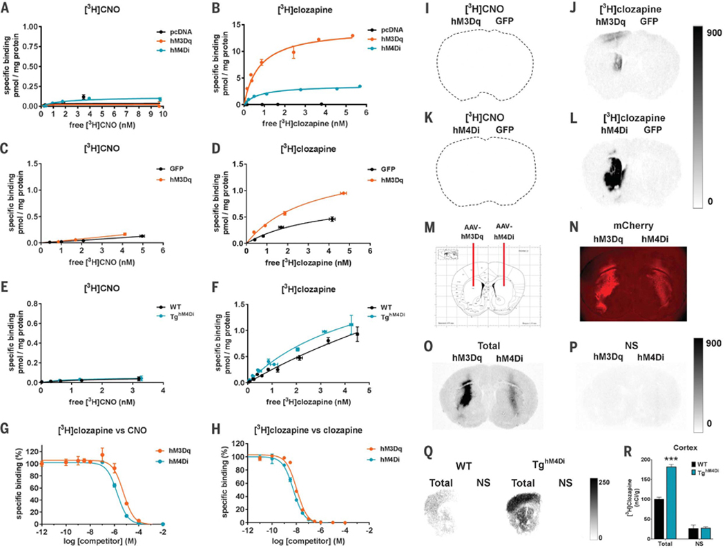
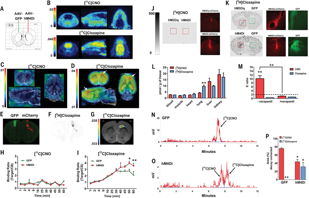
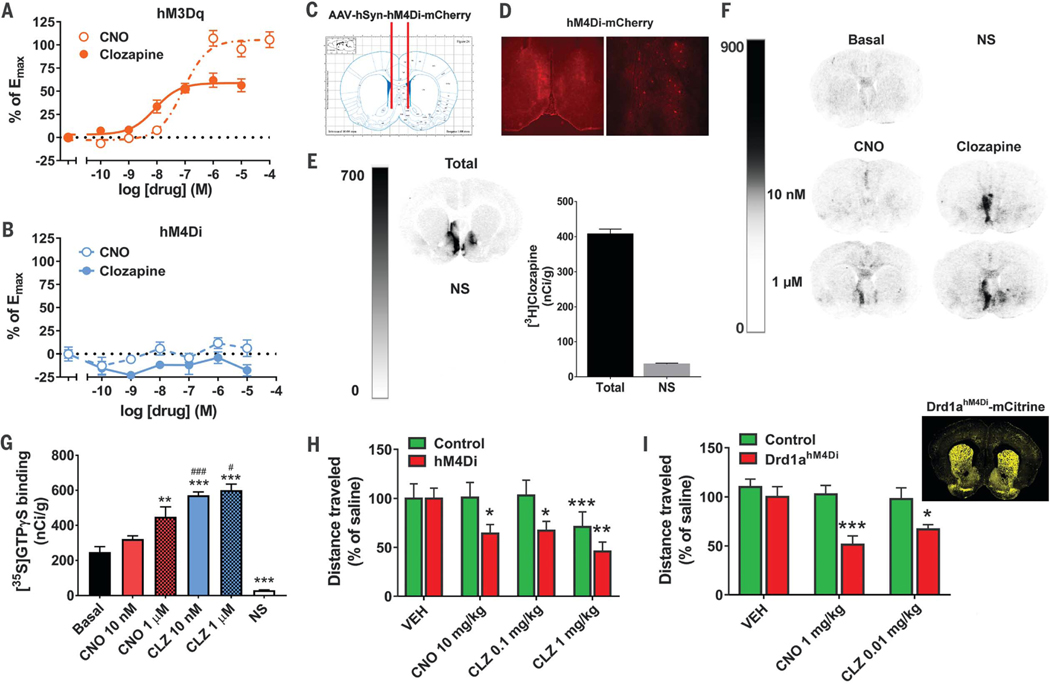
The chemogenetic technology DREADD (designer receptors exclusively activated by designer drugs) is widely used for remote manipulation of neuronal activity in freely moving animals. DREADD technology posits the use of "designer receptors," which are exclusively activated by the "designer drug" clozapine N-oxide (CNO). Nevertheless, the in vivo mechanism of action of CNO at DREADDs has never been confirmed. CNO does not enter the brain after systemic drug injections and shows low affinity for DREADDs. Clozapine, to which CNO rapidly converts in vivo, shows high DREADD affinity and potency. Upon systemic CNO injections, converted clozapine readily enters the brain and occupies central nervous system-expressed DREADDs, whereas systemic subthreshold clozapine injections induce preferential DREADD-mediated behaviors.


Figures

Fig. 1.
Fig. 1.. CNO binds to DREADDs with low affinity, whereas clozapine binds to DREADDs with high affinity.
(A and B) [3H]CNO- or [3H]clozapine-specific binding to HEK-293 membranes expressing DREADDs or pcDNA (control). (C to F) Mouse striatum transduced expressing [(C) and (D)] hM3Dq or GFP (n = 2 mice), or [(E) and (F)] hM4Di-Tg and WT mouse whole-brain tissue (n = 2 mice). (G and H) Competition curves for [3H]clozapine (2.5 nM) against (G) CNO or (H) clozapine in HEK-293 membranes expressing DREADDs. All data points (means ± SEM) are from a representative experiment performed in triplicate. (I to L) [3H]CNO (1 nM) and [3H]clozapine (1 nM) autoradiograms from mice expressing [(I) and (J)] hM3Dq-mCherry or [(K) and (L)] hM4Di-mCherry in the left striatum and GFP in the right striatum (n = 4 mice). (M) AAV injection schematic. (N) Immunofluorescence image showing high DREADD-mCherry expression in dorsal striatum. (O and P) [3H]clozapine (3.5 nM) autoradio-grams from a rat injected with DREADDs as described above showing total and nonspecific (NS) binding. (Q) [3H]clozapine (3.5 nM) autoradiograms showing total and NS binding in hM4Di-Tg and WT mice (n = 2 mice). (R) Densitometric quantification of binding in the cortex of hM4Di-Tg and WT mice. Eight sections from each mouse were mounted onto two slides and assessed for total binding; four sections were mounted onto separate slides and assessed for NS binding. Statistical significance calculated by use of two-way analysis of variance (ANOVA) with genotype and binding condition as factors and Tukey’s post hoc test comparing WT versus hM4Di-Tg mice (***P < 0.001). All values represent mean ± SEM.
Fig. 2.
Fig. 2.. CNO does not cross the BBB while converted clozapine engages DREADDs in vivo.
(A) AAV injection sites. (B) Whole-head PET images (averaged from 1 to 60 min) after intravenous injection of [11C]CNO (n = 2 rats) or [11C]clozapine (n = 2 rats). (C) [11C]CNO or (D) [11C]clozapine PET brain images (averaged from 45 to 60 min) coregistered to a MRI rat template (white arrows indicate hM4Di expression site). (E) Immunohistochemical detection of GFP and hM4Di-mCherry from the rat shown above. (F) [3H]clozapine autoradiogram from an adjacent section to (E). (G) Thresholded [11C]clozapine PET-MRI coregistered image corresponding to (E) and (F). (H) [11C]CNO (n = 2 rats) or (I) [11C]clozapine (n = 2 rats) uptake quantified as target-to-reference (cerebellum) binding ratio, two-way repeated measures ANOVA with time versus group (GFP versus hM4Di) and Sidak’s post hoc test (50 min, P < 0.05; 60 min, P < 0.01). (J and K) Autoradiographic and fluorescent images from mice transduced with hM3Dq-mCherry, hM4Di-mCherry, or GFP in striatum. (L) Organ distribution of [3H]CNO or [3H]clozapine (unpaired Student’s t test). (M) Efflux (E) ratios of CNO and clozapine in the P-gp assay, two-way ANOVA, with drug and condition as factors and Tukey’s post hoc test. (N and O) Representative chromatograms of brain extracts from rats injected with [11C]CNO (n = 4 rats). (P) Percentage of [11C]CNO and converted [11C]clozapine, two-way ANOVA with drug and hemisphere as factors and Tukey’s post hoc test comparing GFP-injected [11C]CNO versus [11C]clozapine (P < 0.01), GFP-[11C]CNO versus hM4Di-[11C]CNO (P < 0.05), and GFP-[11C]clozapine versus hM4Di-[11C]clozapine (P < 0.05, one-tailed). *P < 0.05, **P < 0.01. All values represent mean ± SEM.
Fig. 3.
Fig. 3.. Clozapine potently activates DREADDs and leads to DREADD-specific behavioral responses.
(A and B) Ca2+ dose-response for CNO and clozapine in HEK-293 cells expressing (A) hM3Dq or (B) hM4Di (from four experiments performed in triplicate). (C) Schematic illustration of AAV-hM4Di bilateral injection. (D) Fluorescence microscopy of brain sections from (C). (E) [3H]clozapine autoradiography of total and NS binding in brain sections from the rat in (D). (F and G) [35S]GTPγS autoradiography with adjacent sections treated with vehicle (Basal), CNO, or clozapine, or nonradioactive GTP (NS) illustrated (F) as a panel and (G) after densitometric quantification (n = 6 sections per condition, performed in duplicate). Statistical significance was calculated by means of one-way ANOVA followed by a Sidak post hoc multiple comparison test (**P < 0.01, **P < 0.001 compared with Basal, and #P < 0.05, ###P < 0.001 compared with the respective dose of CNO). (H) Rats expressing hM4Di-mCherry (n = 10 rats) and controls (GFP or sham) (n = 9 rats) in the accumbens/basal forebrain were tested for locomotor activity after injection of vehicle, CNO, and clozapine. Statistical significance was calculated by using repeated measures two-way ANOVA with group (control versus hM4Di) and drug as factors and Tukey’s post hoc multiple comparison test (*P < 0.05, **P < 0.01, ***P < 0.001 compared with the respective vehicle). (I) Drd1ahM4Dl mice (n = 6 or 7) and controls (n = 6 to 9) were injected with vehicle, CNO, or clozapine, and their ambulatory activity was recorded. Statistical significance was calculated by using repeated measures two-way ANOVA with group (control versus Drd1ahM4Di) and drug as factors and Tukey’s post hoc multiple comparison test (*P < 0.05, ***P < 0.001 compared with the respective vehicle). All values represent mean ± SEM.

Comment in

References

    1. Armbruster BN, Li X, Pausch MH, Herlitze S, Roth BL, Proc. Natl. Acad. Sci. U.S.A 104, 5163–5168 (2007). - PMC - PubMed
    1. English JG, Roth BL, JAMA Neurol. 72, 1361–1366 (2015). - PubMed
    1. Urban DJ, Roth BL, Annu. Rev. Pharmacol. Toxicol 55, 399–417 (2015). - PubMed
    1. Ji B et al., J. Neurosci 36, 11544–11558 (2016). - PMC - PubMed
    1. Schotte A et al., Psychopharmacology (Berl.) 124, 57–73 (1996). - PubMed

Publication types